Fraud Blocker Skip to main content

Cerebellum Collection

The cerebellum, often referred to as the "little brain, " is a remarkable structure located at the base of the brain

Background imageCerebellum Collection: Cerebellum tissue, light micrograph

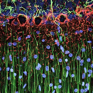
Cerebellum tissue, light micrograph
Cerebellum tissue. Confocal light micrograph of a section through the cerebellum of the brain. Purkinje cells, a type of neuron (nerve cell), are red

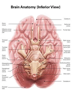
Background imageCerebellum Collection: Anatomy of human brain, inferior view

Anatomy of human brain, inferior view

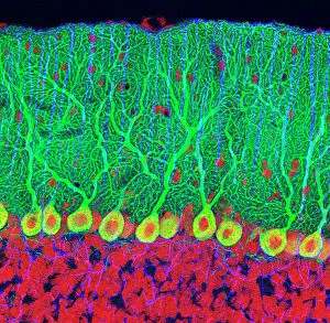
Background imageCerebellum Collection: Purkinje nerve cells in the cerebellum

Purkinje nerve cells in the cerebellum
Purkinje cells in the cerebellum. Fluorescent light micrograph of Purkinje cells (green) in the cerebellum of the brain. Purkinje nerve cells have a flask-like body from which numerous highly

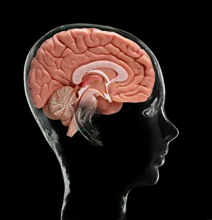
Background imageCerebellum Collection: Human brain, model

Human brain, model. Composite image of a sectioned model human brain inside a glass head. This view shows some of the brains major structures, including: the cerebral cortex (folded areas of tissue)

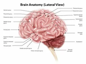
Background imageCerebellum Collection: Human brain anatomy, lateral view

Human brain anatomy, lateral view

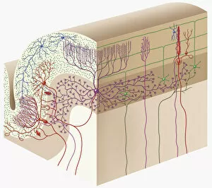
Background imageCerebellum Collection: Cell types in the mammalian cerebellum: drawing, 1894, by the Spanish histologist Santiago Ramon y

Cell types in the mammalian cerebellum: drawing, 1894, by the Spanish histologist Santiago Ramon y Cajal (1852-1934)
NERVE CELLS, 1894. Cell types in the mammalian cerebellum: drawing, 1894, by the Spanish histologist Santiago Ramon y Cajal (1852-1934)

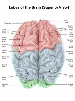
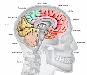
Background imageCerebellum Collection: Superior view of human brain with colored lobes and labels

Superior view of human brain with colored lobes and labels

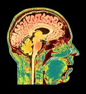
Background imageCerebellum Collection: Brain anatomy, MRI scan

Brain anatomy, MRI scan
Brain anatomy. Coloured magnetic resonance imaging (MRI) scan of the human head from the side. The sagittal scan has halved the brain, revealing the internal anatomy

Background imageCerebellum Collection: Purkinje nerve cells in the cerebellum

Purkinje nerve cells in the cerebellum
Purkinje cells in the cerebellum. Fluorescent light micrograph of Purkinje cells (green) in the cerebellum of the brain. Purkinje nerve cells have a flask-like body from which numerous highly

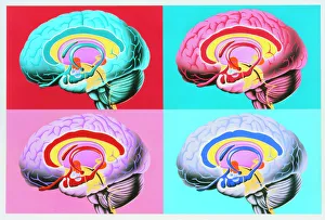
Background imageCerebellum Collection: Artworks showing the limbic system of the brain

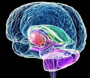
Artworks showing the limbic system of the brain
Limbic system. Four coloured illustrations of the human brain highlighting structures of the limbic system. The limbic system governs activities such as fear, rage, pleasure

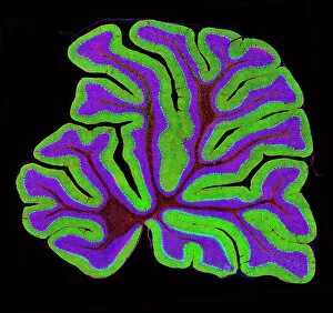
Background imageCerebellum Collection: Cerebellum structure, light micrograph

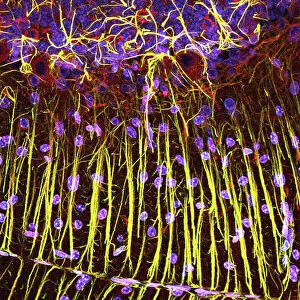
Cerebellum structure, light micrograph
Cerebellum structure. Fluorescent light micrograph of a section through the cerebellum of the brain. The cerebellum comprises three main layers

Background imageCerebellum Collection: Brain anatomy, artwork

Brain anatomy, artwork
Brain anatomy, computer artwork. The cerebellum is purple the corpus callosum is green

Background imageCerebellum Collection: Glial cells, confocal light micrograph

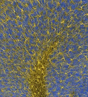
Glial cells, confocal light micrograph
Glial cells. Confocal light micrograph of glial cells from the cerebellum of the brain. Glial cells are nervous system cells that provide structural support and protection for neurons (nerve cells)

Background imageCerebellum Collection: Brain anatomy, artwork

Brain anatomy, artwork
Brain anatomy, computer artwork. The cerebellum is purple the corpus callosum is green

Background imageCerebellum Collection: Brain anatomy, artwork

Brain anatomy, artwork
Brain anatomy, computer artwork. The cerebellum is purple the corpus callosum is green

Background imageCerebellum Collection: Cerebellum tissue, light micrograph

Cerebellum tissue, light micrograph
Cerebellum tissue. Confocal light micrograph of a section through the cerebellum of the brain showing two types of glial cells (support cells); astrocytes (star-shaped)

Background imageCerebellum Collection: Cerebellum tissue, light micrograph

Cerebellum tissue, light micrograph
Cerebellum tissue. Confocal light micrograph of a section through the cerebellum of the brain. Purkinje cells, a type of neuron (nerve cell), are red

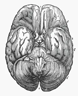
Background imageCerebellum Collection: Under Surface of the Human Brain Engraved Illustration, Circa1880

Under Surface of the Human Brain Engraved Illustration, Circa1880
Beautifully Illustrated Antique Engraved Victorian Illustration of the Under Surface of the Human Brain Engraved Illustration, Circa1880. Source: The Popular Science Monthly, By E.L. and W.J

Background imageCerebellum Collection: Digital illustration of Bocas area (purple), Wernickes area (orange)

Digital illustration of Bocas area (purple), Wernickes area (orange), Geschwinds territory (turquoise) and arcuate fasciculus (green) in human brain

Background imageCerebellum Collection: Conceptual image of human skull and spinal cord

Conceptual image of human skull and spinal cord

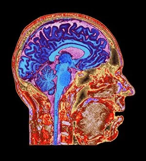
Background imageCerebellum Collection: Normal human brain, MRI scan C016 / 8845

Normal human brain, MRI scan C016 / 8845
Brain. Coloured magnetic resonance imaging (MRI) scan of a sagittal section through a patients head showing a healthy human brain

Background imageCerebellum Collection: Healthy brain, MRI scans

Healthy brain, MRI scans
Healthy brain. Coloured magnetic resonance imaging (MRI) scans through the head of a 55 year old man, showing the brain sectioned in coronal (frontal) view

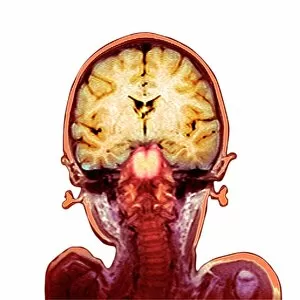
Background imageCerebellum Collection: Childs brain, MRI scan

Childs brain, MRI scan
Childs brain. Coloured magnetic resonance imaging (MRI) scan of an coronal (vertical) section through the head and brain of a healthy 3-year-old child, passing through the ears

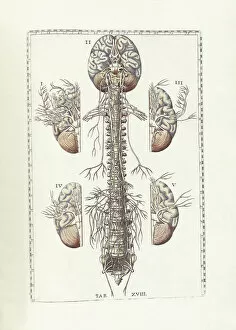
Background imageCerebellum Collection: The science of human anatomy by Bartholomeo Eustachi

The science of human anatomy by Bartholomeo Eustachi, depicting the shape, size and relative position of the organs of the human body. Circa 18th century

Background imageCerebellum Collection: Illustration of bird brain, including cerebrum, cerebellum, and medulla

Illustration of bird brain, including cerebrum, cerebellum, and medulla

Background imageCerebellum Collection: Digital cross section illustration of human cerebellar cortex

Digital cross section illustration of human cerebellar cortex

Background imageCerebellum Collection: Normal human brain, MRI scan C016 / 8850

Normal human brain, MRI scan C016 / 8850
Brain. Coloured magnetic resonance imaging (MRI) scan of a sagittal section through a patients head showing a healthy human brain

Background imageCerebellum Collection: Purkinje nerve cell, TEM C014 / 0583

Purkinje nerve cell, TEM C014 / 0583
Purkinje nerve cell. Transmission electron micrograph (TEM) of a purkinje nerve cell (green) from the cerebellum of the brain, showing the cell body (centre) and its primary dendrite (cell process)

Background imageCerebellum Collection: Brain anatomy

Brain anatomy. Computer artwork of strands of nerve cells (neurons) in front of an exploded view of the brain seen from behind

Background imageCerebellum Collection: Human brain, illustration

Human brain, illustration
Illustration of a cross-section of the brain showing the various lobes. The lobes are shown in different colours - red (frontal), green (parietal), yellow (occipital), orange (temporal)

Background imageCerebellum Collection: Interior of the brain and cerebellum. Copperplate engraving by Robert Benard after an illustration

Interior of the brain and cerebellum. Copperplate engraving by Robert Benard after an illustration by Albrecht von
FLO4611652 Interior of the brain and cerebellum. Copperplate engraving by Robert Benard after an illustration by Albrecht von Haller from Denis Diderot's Encyclopedia, Pellet, Geneva

Background imageCerebellum Collection: Female brain, computer artwork

Female brain, computer artwork
cerebellum:CB2, cerebrum:CB2, spinal column:CB2, adult:CB2, head and shoulders:CB2, side view:CB2, anatomy:CB2, illustration:CB2, black background:CB2, female:CB2, translucent:CB2, nobody:CB2

Background imageCerebellum Collection: Female brain, computer artwork

Female brain, computer artwork
cerebellum:CB2, cerebrum:CB2, spinal column:CB2, head and shoulders:CB2, side view:CB2, anatomy:CB2, illustration:CB2, black background:CB2, female:CB2, translucent:CB2, pituitary gland:CB2

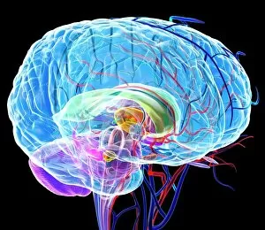
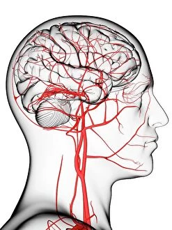
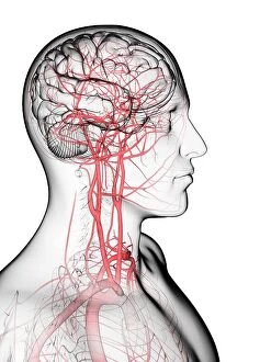
Background imageCerebellum Collection: Brain's blood supply, artwork

Brain's blood supply, artwork
Brain's blood supply, computer artwork.

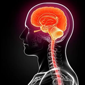
Background imageCerebellum Collection: Male brain, computer artwork

Male brain, computer artwork
cerebellum:CB2, cerebrum:CB2, adult:CB2, head and shoulders:CB2, side view:CB2, visual arts:CB2, anatomy:CB2, illustration:CB2, black background:CB2, male:CB2, translucent:CB2, nobody:CB2

Background imageCerebellum Collection: Brain's blood supply, artwork

Brain's blood supply, artwork
Brain's blood supply, computer artwork.

Background imageCerebellum Collection: Brain's blood supply, artwork

Brain's blood supply, artwork
Brain's blood supply, computer artwork.

Background imageCerebellum Collection: Male brain, computer artwork

Male brain, computer artwork
men, cerebellum, cerebrum, spinal column, head and shoulders, side view, anatomy, illustration, black background, translucent, nobody, human body, brain stem, human representation, male likeness

Background imageCerebellum Collection: Male brain, computer artwork

Male brain, computer artwork
men, cerebellum, cerebrum, spinal column, head and shoulders, side view, anatomy, illustration, black background, translucent, nobody, human body, brain stem, human representation, male likeness

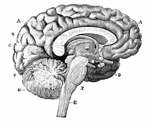
Background imageCerebellum Collection: Antique medical scientific illustration high-resolution: brain

Antique medical scientific illustration high-resolution: brain

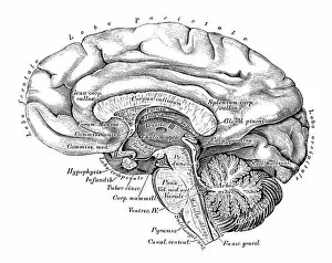
Background imageCerebellum Collection: Human anatomy scientific illustrations: Brain side view

Human anatomy scientific illustrations: Brain side view
Human anatomy scientific illustrations with latin/italian labels: Brain side view

Background imageCerebellum Collection: Human head anatomy, artwork

Human head anatomy, artwork
Human head anatomy, computer artwork

Background imageCerebellum Collection: Human Brain, Lateral View

Human Brain, Lateral View
Lateral view of the human brain showing the cerebral cortex, pons, medulla oblongata, cerebellum and spinal cord

Background imageCerebellum Collection: Birds Nests (engraving)

Birds Nests (engraving)
1096667 Birds Nests (engraving) by English School, (19th century); Private Collection; (add.info.: Birds Nests. Illustration for The Illustrated London News)

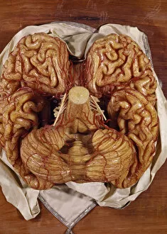
Background imageCerebellum Collection: Model of a brain (wax)

Model of a brain (wax)
BEN360191 Model of a brain (wax) by Susini, Clemente (1754-1814); Museo della Specola, Florence, Italy; (add.info.: cervello; scultura di cera; ); Photo eRaffaello Bencini; Italian, out of copyright

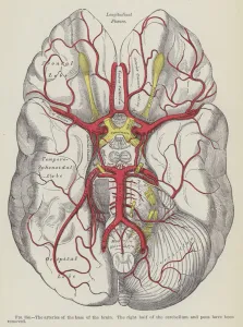
Background imageCerebellum Collection: The arteries of the base of the brain (engraving)

The arteries of the base of the brain (engraving)
3634191 The arteries of the base of the brain (engraving) by English School, (19th century); Private Collection; (add.info.: The arteries of the base of the brain)

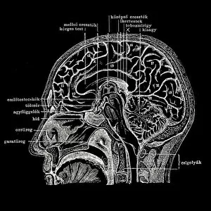
Background imageCerebellum Collection: Human head section

Human head section
Longitudinal section of the human head

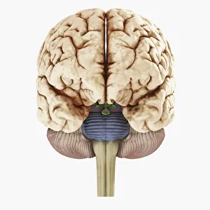
Background imageCerebellum Collection: Digital illustration of showing front view of human brain

Digital illustration of showing front view of human brain


All Professionally Made to Order for Quick Shipping

The cerebellum, often referred to as the "little brain, " is a remarkable structure located at the base of the brain. This intricate tissue plays a crucial role in coordinating movement, balance, and posture. When examined under a light micrograph, the cerebellum reveals its complex network of cells and fibers that work together seamlessly. From an inferior view of the human brain anatomy model, one can appreciate how the cerebellum fits snugly beneath the cerebral hemispheres. Its distinct lobes are clearly labeled and color-coded in a superior view of the brain. This lateral perspective allows us to understand its position relative to other regions within this vital organ. Intriguingly, Spanish histologist Santiago Ramon y Cajal's 1894 drawing showcases various cell types found within mammalian cerebellum tissue. His meticulous illustrations provide valuable insights into our understanding of this fascinating part of our brains. Glial cells are another essential component highlighted in confocal light micrographs. These support cells play a critical role in maintaining neuronal health and function within the cerebellum. Purkinje nerve cells stand out prominently when examining both artwork depicting them and actual images from MRI scans or light microscopy. These specialized neurons serve as key players in transmitting signals throughout this region of our brains. Artworks showcasing different aspects of brain anatomy further emphasize just how interconnected all these structures are within our limbic system—the emotional center responsible for memory formation and regulation among other functions.