Fraud Blocker Skip to main content

Cerebellum Collection (page 3)

The cerebellum, often referred to as the "little brain, " is a remarkable structure located at the base of the brain

Background imageCerebellum Collection: Digital illustration of head in profile showing dorsal and ventral pathways of brain

Digital illustration of head in profile showing dorsal and ventral pathways of brain

Background imageCerebellum Collection: Digital illustration of domestic cat showing cerebellum highlighted in orange

Digital illustration of domestic cat showing cerebellum highlighted in orange

Background imageCerebellum Collection: Digital illustration of highlighted areas in human brain affected by motor disorders

Digital illustration of highlighted areas in human brain affected by motor disorders

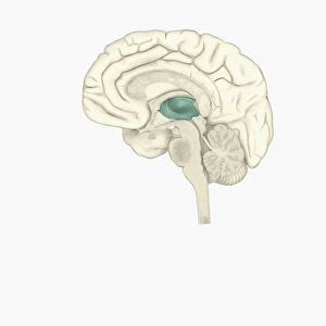
Background imageCerebellum Collection: Digital illustration of thalamus in human brain highlighted in green

Digital illustration of thalamus in human brain highlighted in green

Background imageCerebellum Collection: Digital illustration of insula in human brain highlighted in red

Digital illustration of insula in human brain highlighted in red

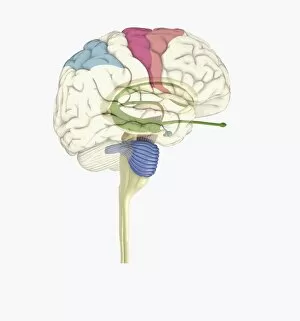
Background imageCerebellum Collection: Digital illustration of head in profile showing motor cortex (pink)

Digital illustration of head in profile showing motor cortex (pink), frontal area and amygdala (green), auditory cortex (orange), and visual cortex in brain

Background imageCerebellum Collection: Digital illustration of head in profile showing visual cortex (blue), motor cortex (pink)

Digital illustration of head in profile showing visual cortex (blue), motor cortex (pink), auditory cortex (orange), frontal area and amygdala (green) brain

Background imageCerebellum Collection: Digital illustration of head in profile showing frontal lobe and temporal lobe in brain

Digital illustration of head in profile showing frontal lobe and temporal lobe in brain

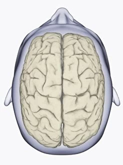
Background imageCerebellum Collection: Digital illustration of head showing left and right areas of brain seen from above

Digital illustration of head showing left and right areas of brain seen from above

Background imageCerebellum Collection: Digital illustration of left hemisphere (yellow) and right hemisphere (blue) of human brain

Digital illustration of left hemisphere (yellow) and right hemisphere (blue) of human brain

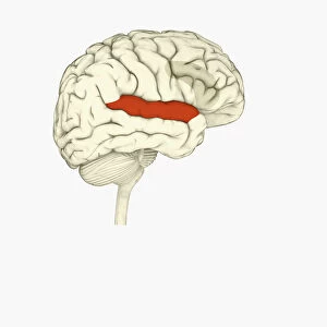
Background imageCerebellum Collection: Digital illustration of superior temporal sulcas (red), orbitofrontal cortex and amygdala (green)

Digital illustration of superior temporal sulcas (red), orbitofrontal cortex and amygdala (green), in human brain

Background imageCerebellum Collection: Digital illustration of posterior cingulate cortex (blue), medial frontal gyrus (yellow)

Digital illustration of posterior cingulate cortex (blue), medial frontal gyrus (yellow), and orbitofrontal prefrontal cortex (green) in human brain

Background imageCerebellum Collection: Digital illustration of parietal lobe (green), posterior superior temporal sulcas (blue)

Digital illustration of parietal lobe (green), posterior superior temporal sulcas (blue), temperal pole (grey), dorsolateral prefrontal cortex, amygdala

Background imageCerebellum Collection: Digital illustration of emotional response areas in human brain

Digital illustration of emotional response areas in human brain

Background imageCerebellum Collection: Digital illustration of anterior cingulate cortex (grey), and medial frontal cortex (green)

Digital illustration of anterior cingulate cortex (grey), and medial frontal cortex (green) in human brain

Background imageCerebellum Collection: Digital illustration of human brain

Digital illustration of human brain

Background imageCerebellum Collection: Digital illustration of reward pathway in human brain

Digital illustration of reward pathway in human brain

Background imageCerebellum Collection: Digital illustration of human brain associated with full awareness

Digital illustration of human brain associated with full awareness

Background imageCerebellum Collection: Digital illustration of areas of information highlighted in human brain

Digital illustration of areas of information highlighted in human brain

Background imageCerebellum Collection: Digital illustration of human brain with orbitofrontal cortex and amygdala highlighted in green

Digital illustration of human brain with orbitofrontal cortex and amygdala highlighted in green

Background imageCerebellum Collection: Digital illustration of head in profile showing mirror neurons in human brain highlighted in

Digital illustration of head in profile showing mirror neurons in human brain highlighted in purple, pink and green

Background imageCerebellum Collection: Digital illustration of head in profile showing area of mirror neuron in brain highlighted in orange

Digital illustration of head in profile showing area of mirror neuron in brain highlighted in orange

Background imageCerebellum Collection: Digital illustration of frontal lobe and parietal lobe areas (orange) in left hemispheres (blue)

Digital illustration of frontal lobe and parietal lobe areas (orange) in left hemispheres (blue), and pathway of data from parietal lobe to frontal lobe (green) in human brain

Background imageCerebellum Collection: Digital illustration of right superior temporal sulcas, and anterior cingulate cortex highlighted

Digital illustration of right superior temporal sulcas, and anterior cingulate cortex highlighted in red and grey in human brain

Background imageCerebellum Collection: Digital illustration of anterior insular, anterior cingulate cortex

Digital illustration of anterior insular, anterior cingulate cortex, and ventromedial prefrontal cortex highlighted in human brain

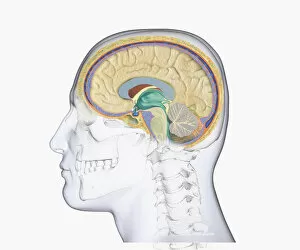
Background imageCerebellum Collection: Digital illustration of head in profile showing skull, brain, and spine

Digital illustration of head in profile showing skull, brain, and spine

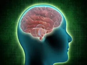
Background imageCerebellum Collection: Digital illustration of head in profile showing brain

Digital illustration of head in profile showing brain

Background imageCerebellum Collection: Digital illustration of human brain with primary auditory cortex highlighted in orange and red

Digital illustration of human brain with primary auditory cortex highlighted in orange and red

Background imageCerebellum Collection: Digital illustration of head in profile showing memory areas of brain

Digital illustration of head in profile showing memory areas of brain

Background imageCerebellum Collection: Digital illustration of head in profile showing bundle of nerve fibres connecti ng Brocas area

Digital illustration of head in profile showing bundle of nerve fibres connecti ng Brocas area and Wernickes area in human brain

Background imageCerebellum Collection: Digital illustration of areas associated with memory in human brain

Digital illustration of areas associated with memory in human brain

Background imageCerebellum Collection: Digital illustration of hippocampus (green) and pons (blue) in left hemisphere of human brain

Digital illustration of hippocampus (green) and pons (blue) in left hemisphere of human brain

Background imageCerebellum Collection: Digital illustration of human brain showing frontal cortex in green

Digital illustration of human brain showing frontal cortex in green

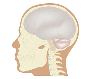
Background imageCerebellum Collection: Cross section biomedical illustration of the brain and skull at 18 years of age

Cross section biomedical illustration of the brain and skull at 18 years of age

Background imageCerebellum Collection: Cross section biomedical illustration of voluntary pathway of human brain

Cross section biomedical illustration of voluntary pathway of human brain

Background imageCerebellum Collection: Cross section biomedical illustration of brain and autonomic pathways in the autonomic nervous

Cross section biomedical illustration of brain and autonomic pathways in the autonomic nervous system

Background imageCerebellum Collection: Cross section biomedical illustration of spinal reflex pathway of human brain

Cross section biomedical illustration of spinal reflex pathway of human brain

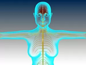
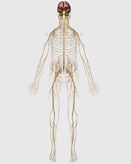
Background imageCerebellum Collection: Conceptual image of female nervous system with brain

Conceptual image of female nervous system with brain

Background imageCerebellum Collection: Conceptual image of female body with brain, side view

Conceptual image of female body with brain, side view

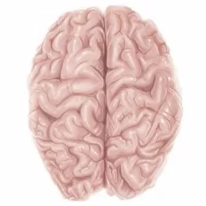
Background imageCerebellum Collection: Superior view of human brain

Superior view of human brain

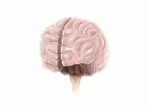
Background imageCerebellum Collection: Brain surface anatomy

Brain surface anatomy

Background imageCerebellum Collection: Sciatica caused from herniated disc

Sciatica caused from herniated disc

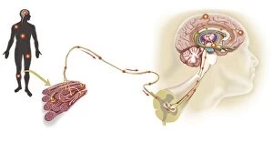
Background imageCerebellum Collection: Pathway of a pain message via sensory nerve in injured muscle

Pathway of a pain message via sensory nerve in injured muscle, to pain gate in spinal cord to limbic system, frontal cortex and sensory cortex in the human brain

Background imageCerebellum Collection: Medical illustration of peripheral nervous system with brain

Medical illustration of peripheral nervous system with brain

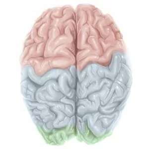
Background imageCerebellum Collection: Superior view of human brain with colored lobes

Superior view of human brain with colored lobes

Background imageCerebellum Collection: Anatomy of pituitary gland

Anatomy of pituitary gland

Background imageCerebellum Collection: Representation of how our senses affect our thoughts

Representation of how our senses affect our thoughts

Background imageCerebellum Collection: Conceptual image of human brain with neurons

Conceptual image of human brain with neurons in background


All Professionally Made to Order for Quick Shipping

The cerebellum, often referred to as the "little brain, " is a remarkable structure located at the base of the brain. This intricate tissue plays a crucial role in coordinating movement, balance, and posture. When examined under a light micrograph, the cerebellum reveals its complex network of cells and fibers that work together seamlessly. From an inferior view of the human brain anatomy model, one can appreciate how the cerebellum fits snugly beneath the cerebral hemispheres. Its distinct lobes are clearly labeled and color-coded in a superior view of the brain. This lateral perspective allows us to understand its position relative to other regions within this vital organ. Intriguingly, Spanish histologist Santiago Ramon y Cajal's 1894 drawing showcases various cell types found within mammalian cerebellum tissue. His meticulous illustrations provide valuable insights into our understanding of this fascinating part of our brains. Glial cells are another essential component highlighted in confocal light micrographs. These support cells play a critical role in maintaining neuronal health and function within the cerebellum. Purkinje nerve cells stand out prominently when examining both artwork depicting them and actual images from MRI scans or light microscopy. These specialized neurons serve as key players in transmitting signals throughout this region of our brains. Artworks showcasing different aspects of brain anatomy further emphasize just how interconnected all these structures are within our limbic system—the emotional center responsible for memory formation and regulation among other functions.