Mouse Mat > Animals > Fishes > Related Images
Mouse Mat : Fish embryo, artwork


![]()
Home Decor from Science Photo Library
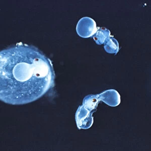
Fish embryo, artwork
Fish embryo. Historical artwork showing stages in the development of a fish embryo. Figures 1 and 2 show gastrulation, the process of differentiation whereby the 3 germ layers (ectoderm, mesoderm and endoderm) are formed. Figure 3 shows neuralation, the formation of the neural tube, which will become the brain and spinal cord. Figure 4 shows the somites (blocks of mesoderm on either sode of neural tube), which will give rise to muscular and skeletal tissues. Figure 5 shows further differentiation of the head
Science Photo Library features Science and Medical images including photos and illustrations
Media ID 6303105
© MEHAU KULYK/SCIENCE PHOTO LIBRARY
Developing Development Developmental Biology Differentiation Ectoderm Embryo Embryology Endoderm Formation Forming Gastrula Gastrulation History Of Science Invagination Neural Tube Poster Somites Stage Stages Vertebrate Vertebrates Mesoderm Nervous System
Mouse Mat
A high quality photographic print manufactured into a durable wipe clean mouse mat (27x22cm) with a non slip backing, which works with all mice.
Archive quality photographic print in a durable wipe clean mouse mat with non slip backing. Works with all computer mice
Estimated Image Size (if not cropped) is 17.5cm x 25.4cm (6.9" x 10")
Estimated Product Size is 21.8cm x 26.9cm (8.6" x 10.6")
These are individually made so all sizes are approximate
Artwork printed orientated as per the preview above, with portrait (vertical) orientation to match the source image.
EDITORS COMMENTS
This print captures the intricate stages of development in a fish embryo, showcasing the remarkable process of differentiation. The historical artwork showcases five key figures that depict various milestones in embryonic growth. Figures 1 and 2 beautifully illustrate gastrulation, a critical phase where three germ layers - ectoderm, mesoderm, and endoderm - are formed. This process lays the foundation for the formation of different tissues and organs within the developing fish. Figure 3 focuses on neuralation, highlighting the formation of the neural tube which will eventually develop into both the brain and spinal cord. This crucial step sets the stage for a well-developed nervous system. Figure 4 unveils somites, blocks of mesoderm found on either side of the neural tube. These somites hold immense potential as they give rise to muscular and skeletal tissues, shaping not only physical structure but also mobility. Lastly, Figure 5 portrays further differentiation specifically within the head region. It showcases how this complex organism continues to evolve with each passing stage. This mesmerizing artwork provides an invaluable glimpse into developmental biology's rich history while shedding light on vertebrate embryology. It serves as a testament to our fascination with life's intricate processes and highlights how science has unraveled nature's secrets over time.
MADE IN THE UK
Safe Shipping with 30 Day Money Back Guarantee
FREE PERSONALISATION*
We are proud to offer a range of customisation features including Personalised Captions, Color Filters and Picture Zoom Tools
SECURE PAYMENTS
We happily accept a wide range of payment options so you can pay for the things you need in the way that is most convenient for you
* Options may vary by product and licensing agreement. Zoomed Pictures can be adjusted in the Basket.