Mounted Print > Animals > Mammals > Muridae > Water Mouse
Mounted Print : Xylem and parenchyma in Rhubarb stem


![]()
Mounted Prints from Science Photo Library
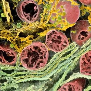
Xylem and parenchyma in Rhubarb stem
Rhubarb stem. Coloured scanning electron micrograph (SEM) of a longitudinal section through a rhubarb stem, Rheum rhaponticum. Cut xylem vessels are coloured brown, and parenchyma cells are coloured green. Xylem vessels are responsible for the upward transport of water and solutes in the plant, from the roots into the stem and leaves. Here, these vessels are reinforced and strengthened with spiral bands of lignin. Spiral bands allow xylem vessels to elongate and grow lengthwise. Parenchyma cells form a ground tissue in which other tissues, such as xylem, are embedded. Magnification: x290 at 5x7cm size. x1000 at 10x8ins
Science Photo Library features Science and Medical images including photos and illustrations
Media ID 9194231
© POWER AND SYRED/SCIENCE PHOTO LIBRARY
Parenchyma Plants Stem Tracheid Vessel Xylem
10"x8" Mount with 8"x6" Print
Discover the intricacies of nature with Media Storehouse's Mounted Photos. This captivating image showcases the intricate structure of a Rhubarb stem, revealing the beauty of its xylem and parenchyma cells. Coloured Scanning Electron Micrograph (SEM) by Science Photo Library provides a detailed and vibrant view, perfect for scientific research, educational purposes, or simply to appreciate the wonders of the natural world. Add this stunning mounted photo to your collection and ignite curiosity and inspiration.
Printed on 8"x6" paper and suitable for use in a 10"x8" frame (frame not included). Prints are mounted with card both front and back. Featuring a custom cut aperture to match chosen image. Professional 234gsm Fujifilm Crystal Archive DP II paper.
Photo prints supplied in custom cut card mount ready for framing
Estimated Image Size (if not cropped) is 20.3cm x 15.2cm (8" x 6")
Estimated Product Size is 25.4cm x 20.3cm (10" x 8")
These are individually made so all sizes are approximate
Artwork printed orientated as per the preview above, with landscape (horizontal) orientation to match the source image.
FEATURES IN THESE COLLECTIONS
> Animals
> Mammals
> Muridae
> Water Mouse
EDITORS COMMENTS
This print showcases the intricate structure of a rhubarb stem, revealing its xylem and parenchyma tissues. In this coloured scanning electron micrograph (SEM), we are granted a close-up view of a longitudinal section through the stem of Rheum rhaponticum, commonly known as rhubarb. The brown-coloured cut xylem vessels take center stage in this image, representing their crucial role in transporting water and solutes from the roots to the rest of the plant. Reinforced with spiral bands of lignin, these vessels gain strength and flexibility for elongation and growth along their length. Contrasting against the xylem vessels are vibrant green parenchyma cells that form a ground tissue within which other important tissues like xylem reside. Acting as support cells, they provide structural integrity to the stem while allowing space for essential transport systems. At 290 times magnification on a 5x7cm print or an impressive 1000 times magnification at 10x8ins size, this image captures nature's remarkable complexity at a microscopic level. It serves as a testament to both botanical beauty and scientific curiosity. Courtesy of Science Photo Library, this photograph offers botany enthusiasts an opportunity to appreciate the wonders hidden within plants while reminding us all of nature's awe-inspiring intricacy.
MADE IN THE UK
Safe Shipping with 30 Day Money Back Guarantee
FREE PERSONALISATION*
We are proud to offer a range of customisation features including Personalised Captions, Color Filters and Picture Zoom Tools
SECURE PAYMENTS
We happily accept a wide range of payment options so you can pay for the things you need in the way that is most convenient for you
* Options may vary by product and licensing agreement. Zoomed Pictures can be adjusted in the Basket.