Mounted Print : Section through a rat embryo


![]()
Mounted Prints from Science Photo Library
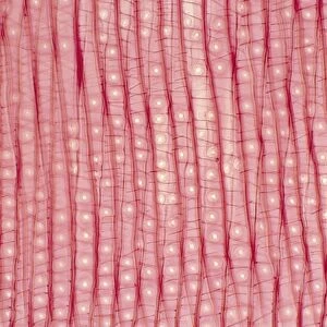
Section through a rat embryo
Light micrograph of a section through a laboratory rat embryo. Between the rows of blue dots (the ribcage) lies the lungs (purple fan shape) and below that the heart (orange round shape). Magnification: x1 at 35mm
Science Photo Library features Science and Medical images including photos and illustrations
Media ID 9306189
© POWER AND SYRED/SCIENCE PHOTO LIBRARY
10"x8" Mount with 8"x6" Print
Discover the intricacies of life with Media Storehouse's Mounted Photos. This captivating image, sourced from Science Photo Library, offers a fascinating glimpse into the developmental stages of a rat embryo. Witness the delicate arrangement of the ribcage (blue dots) and the developing lungs (purple fan shape), with the heart (orange round shape) situated below. A must-have for biology enthusiasts, educators, and researchers, these high-quality mounted photos bring the wonders of science right to your doorstep.
Printed on 8"x6" paper and suitable for use in a 10"x8" frame (frame not included). Prints are mounted with card both front and back. Featuring a custom cut aperture to match chosen image. Professional 234gsm Fujifilm Crystal Archive DP II paper.
Photo prints supplied in custom cut card mount ready for framing
Estimated Image Size (if not cropped) is 13.5cm x 20.3cm (5.3" x 8")
Estimated Product Size is 20.3cm x 25.4cm (8" x 10")
These are individually made so all sizes are approximate
Artwork printed orientated as per the preview above, with portrait (vertical) orientation to match the source image.
EDITORS COMMENTS
This print showcases a remarkable section through a laboratory rat embryo, providing us with an extraordinary glimpse into the intricate world of embryonic development. The image reveals an astonishing level of detail, allowing us to observe the delicate structures that form within the tiny body of this developing rat. The rows of vibrant blue dots represent the ribcage, serving as a protective framework for the growing organs. Nestled between these rows lies a mesmerizing purple fan shape, which represents the lungs taking shape and preparing for their vital role in respiration. Just below them, we can spot an enchanting orange round shape - none other than the heart itself - beating rhythmically even at this early stage. With its magnification set at x1 and captured using light microscopy techniques, this photograph truly captures nature's artistry on a microscopic scale. It serves as a testament to both scientific curiosity and technological advancement in studying animal anatomy. As we gaze upon this awe-inspiring image from Science Photo Library, let our minds wander into contemplation about life's beginnings and marvel at how such complex organisms develop from mere cells. This visual masterpiece invites us to appreciate not only the beauty inherent in every living creature but also highlights humanity's relentless pursuit of knowledge in unraveling nature's mysteries.
MADE IN THE UK
Safe Shipping with 30 Day Money Back Guarantee
FREE PERSONALISATION*
We are proud to offer a range of customisation features including Personalised Captions, Color Filters and Picture Zoom Tools
SECURE PAYMENTS
We happily accept a wide range of payment options so you can pay for the things you need in the way that is most convenient for you
* Options may vary by product and licensing agreement. Zoomed Pictures can be adjusted in the Basket.