Mounted Print : Section through leaf of Zinnia


![]()
Mounted Prints from Science Photo Library
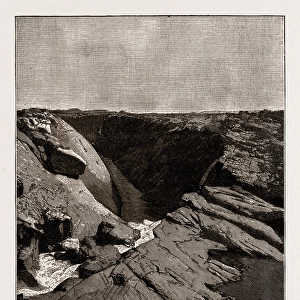
Section through leaf of Zinnia
False-colour scanning electron micrograph of the under surface of a leaf, Zinnia elegans, showing a part of the epidermis removed to reveal the spongy mesophyll layer (left) under the surface. The epidermis (right) consistis of large, irregular cells with waterproofing constituents in the wall. The jigsaw effect corresponds to the epidermal cells. The epidermis shows stomatal pores, which regulate the exchange of air between the atmosphere and the leafs interior. Mesophyll cells are large and attached at limited points only, creating airspaces through which the air entering via the pores circulates freely through the leaf. Magnification: x150 at 6x4.5cm size
Science Photo Library features Science and Medical images including photos and illustrations
Media ID 6289569
© DR JEREMY BURGESS/SCIENCE PHOTO LIBRARY
Epidermis Pore Pores Stoma Stomata Zinnia Elegans
10"x8" Mount with 8"x6" Print
Discover the intricacy of nature with Media Storehouse's Mounted Photos. This captivating image showcases a false-colour scanning electron micrograph of a Zinnia elegans leaf, revealing the mesmerizing structure of its spongy mesophyll layer. Delve deeper into the world of science with our high-quality, mounted photos, perfect for educational displays or personal collections. Experience the beauty of nature in unparalleled detail.
Printed on 8"x6" paper and suitable for use in a 10"x8" frame (frame not included). Prints are mounted with card both front and back. Featuring a custom cut aperture to match chosen image. Professional 234gsm Fujifilm Crystal Archive DP II paper.
Photo prints supplied in custom cut card mount ready for framing
Estimated Image Size (if not cropped) is 20.1cm x 15.2cm (7.9" x 6")
Estimated Product Size is 25.4cm x 20.3cm (10" x 8")
These are individually made so all sizes are approximate
Artwork printed orientated as per the preview above, with landscape (horizontal) orientation to match the source image.
EDITORS COMMENTS
This print offers a mesmerizing glimpse into the intricate world of plant anatomy. A section through the leaf of Zinnia elegans is beautifully captured in this false-colour scanning electron micrograph, revealing its hidden secrets. The under surface of the leaf showcases the spongy mesophyll layer, which appears like a delicate web of interconnected cells. On the right side, we observe the epidermis consisting of large irregular cells with waterproofing constituents in their walls. These unique structures create a jigsaw effect that adds to the visual allure. Notably, within this epidermal layer, stomatal pores are visible - tiny openings that play a crucial role in regulating air exchange between the atmosphere and the interior of leaves. The mesophyll cells on display are noticeably larger and only attached at limited points, resulting in airspaces throughout the leaf. This ingenious design allows for free circulation of air entering via these stomatal pores. It's fascinating to witness nature's ingenuity at work as it ensures efficient gas exchange within plants. With a magnification level set at x150 and presented in a 6x4.5cm size format, this photograph truly captures both scientific precision and artistic beauty simultaneously. Whether you're an avid botany enthusiast or simply appreciate nature's wonders, this image is sure to captivate your imagination and deepen your appreciation for Earth's incredible biodiversity.
MADE IN THE UK
Safe Shipping with 30 Day Money Back Guarantee
FREE PERSONALISATION*
We are proud to offer a range of customisation features including Personalised Captions, Color Filters and Picture Zoom Tools
SECURE PAYMENTS
We happily accept a wide range of payment options so you can pay for the things you need in the way that is most convenient for you
* Options may vary by product and licensing agreement. Zoomed Pictures can be adjusted in the Basket.