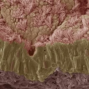
Mounted Print : F. colour SEM of oocysts on stomach of Anopheles


![]()
Mounted Prints from Science Photo Library
F. colour SEM of oocysts on stomach of Anopheles
False-colour scanning electron micrograph of the stomach wall of a mosquito Anopholes stephansii infected with malarial parasites Plasmodium. The small rounded objects covering the outside stomach wall are oocysts, a stage in the life cycle of the parasite. The oocysts grow in size & the contents divide to form sporozoites, which are dispersed through the body cavity when the oocyst bursts. The released sporozoites eventually reach the salivary gland of the mosquito & are transmitted to humans when the female takes a blood meal
Science Photo Library features Science and Medical images including photos and illustrations
Media ID 6468785
© LONDON SCHOOL OF HYGIENE & TROPICAL MEDICINE/SCIENCE PHOTO LIBRARY
Malaria Malarial Parasite Plasmodium Protozoa Protozoan
10"x8" Mount with 8"x6" Print
Discover the intricacies of nature with Media Storehouse's Mounted Photos. This captivating image, sourced from Science Photo Library, showcases a False-colour Scanning Electron Micrograph (SEM) of the stomach wall of an Anopheles stephansii mosquito, infected with malarial parasites Plasmodium. Witness the vibrant colours that reveal the complex structures of the oocysts on the mosquito's stomach, providing a unique glimpse into the life cycle of this deadly parasite. Enhance your scientific research, educational materials, or display with this stunning, high-resolution mounted photo.
Printed on 8"x6" paper and suitable for use in a 10"x8" frame (frame not included). Prints are mounted with card both front and back. Featuring a custom cut aperture to match chosen image. Professional 234gsm Fujifilm Crystal Archive DP II paper.
Photo prints supplied in custom cut card mount ready for framing
Estimated Image Size (if not cropped) is 20.3cm x 14.5cm (8" x 5.7")
Estimated Product Size is 25.4cm x 20.3cm (10" x 8")
These are individually made so all sizes are approximate
Artwork printed orientated as per the preview above, with landscape (horizontal) orientation to match the source image.
EDITORS COMMENTS
This print offers a mesmerizing glimpse into the intricate world of nature's hidden battles. The false-color scanning electron micrograph showcases the stomach wall of an Anopheles mosquito, specifically Anopholes stephansii, infected with malarial parasites known as Plasmodium. The image reveals small rounded objects called oocysts that densely cover the exterior of the mosquito's stomach wall. These oocysts represent a crucial stage in the life cycle of the malaria parasite. As they grow in size, their contents divide to form sporozoites - tiny organisms responsible for transmitting malaria. When these oocysts burst open, countless sporozoites are released into the body cavity of the mosquito. Over time, these resilient creatures navigate their way towards the salivary gland. Once there, they patiently await their next opportunity to infect unsuspecting humans when a female mosquito takes her blood meal. This photograph not only highlights nature's complexity but also serves as a reminder of how delicate our ecosystem truly is. It sheds light on one of humanity's greatest challenges - combating malaria and its devastating impact on millions worldwide. Through this stunning visual representation captured by Science Photo Library, we gain insight into both scientific marvels and urgent global health concerns surrounding this deadly parasitic disease.
MADE IN THE UK
Safe Shipping with 30 Day Money Back Guarantee
FREE PERSONALISATION*
We are proud to offer a range of customisation features including Personalised Captions, Color Filters and Picture Zoom Tools
SECURE PAYMENTS
We happily accept a wide range of payment options so you can pay for the things you need in the way that is most convenient for you
* Options may vary by product and licensing agreement. Zoomed Pictures can be adjusted in the Basket.