
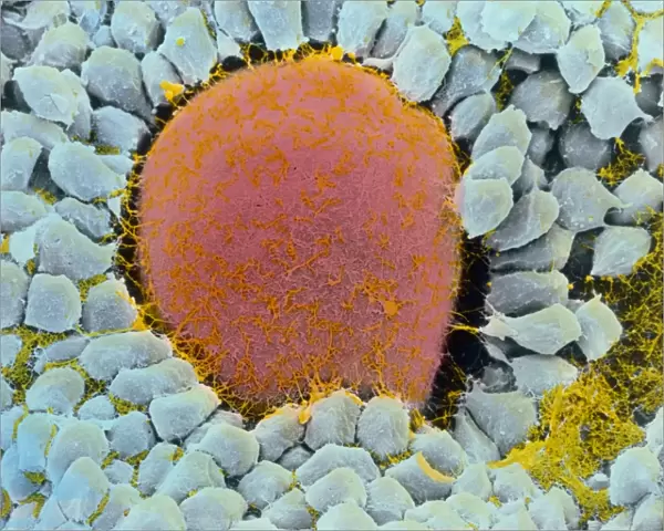
Metal Print : Coloured SEM of egg cell in secondary follicle


![]()
Metal Prints from Science Photo Library
Coloured SEM of egg cell in secondary follicle
Science Photo Library features Science and Medical images including photos and illustrations
Media ID 6455671
© PROFESSOR P.M. MOTTA, G. MACCHIARELLI, S.A NOTTOLA/SCIENCE PHOTO LIBRARY
Egg Cell Female Reproductive System Follicle Oocyte Ovarian Ovum Re Production Secondary Secondary Follicle Secondary Oocyte
20"x16" (51x41cm) Metal Print
Discover the intricacy of life with our Media Storehouse Metal Prints featuring this stunning Coloured SEM of an egg cell in a secondary follicle from Science Photo Library. Each print is meticulously crafted using high-quality metal materials, ensuring vibrant colors and exceptional detail that bring the beauty of science to your home or office. This unique and captivating image provides a glimpse into the complex world of biology, making it an excellent conversation starter and a beautiful addition to any space. Order now and bring the wonders of science into your daily life.
Your image is printed photographically and bonded to a 3.5mm thick, Dibond board (black polyethylene sandwiched between two sheets of white coated aluminium). The panel is then sealed with a gloss protective covering. Supplied complete with a wall mount which holds the print 10mm from the wall.
Made with durable metal and luxurious printing techniques, metal prints bring images to life and add a modern touch to any space
Estimated Product Size is 50.8cm x 40.6cm (20" x 16")
These are individually made so all sizes are approximate
Artwork printed orientated as per the preview above, with landscape (horizontal) or portrait (vertical) orientation to match the source image.
EDITORS COMMENTS
This print from Science Photo Library showcases the intricate beauty of a coloured scanning electron microscope (SEM) image of an egg cell in a secondary follicle. The image provides a fascinating glimpse into the world of reproduction and anatomy within the female reproductive system. The vibrant colours bring to life the various components involved in this crucial process. At its center, we see the secondary oocyte, also known as an ovum or egg cell, surrounded by granulosa cells that play a vital role in nurturing and supporting its development. These granulosa cells form part of the follicular antrum, which can be observed as small spaces filled with fluid surrounding the egg cell. This visual representation offers valuable insights into human fertility and highlights the complexity behind conception. It serves as a reminder of how remarkable our bodies are at creating new life. Science Photo Library has once again captured an awe-inspiring moment through their lens, showcasing both scientific precision and artistic flair. This photograph is not only visually stunning but also serves as a powerful educational tool for those interested in understanding more about reproductive biology.
MADE IN THE UK
Safe Shipping with 30 Day Money Back Guarantee
FREE PERSONALISATION*
We are proud to offer a range of customisation features including Personalised Captions, Color Filters and Picture Zoom Tools
SECURE PAYMENTS
We happily accept a wide range of payment options so you can pay for the things you need in the way that is most convenient for you
* Options may vary by product and licensing agreement. Zoomed Pictures can be adjusted in the Basket.