
Secondary Follicle Collection
A secondary follicle, as seen through various microscopic imaging techniques such as SEM (Scanning Electron Microscopy), TEM (Transmission Electron Microscopy


All Professionally Made to Order for Quick Shipping
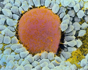
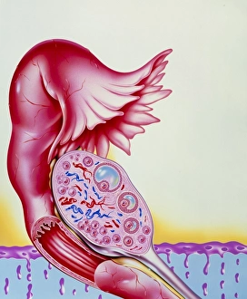
A secondary follicle, as seen through various microscopic imaging techniques such as SEM (Scanning Electron Microscopy), TEM (Transmission Electron Microscopy), and light micrographs, provides a fascinating glimpse into the intricate world of ovarian follicles. In an ovarian follicle, the primary stage is followed by the formation of a secondary follicle. This developmental process can be observed in stunning detail under SEM, where delicate structures are revealed with remarkable clarity. The coloured SEM image showcases an egg cell within a secondary follicle - a crucial stage in reproductive biology. Light micrographs further enhance our understanding of these dynamic structures. They capture the beauty of ovarian follicles at different stages, highlighting their complexity and importance in female fertility. From early development to maturity, each image tells a unique story about ovum growth within a woman's ovary. The TEM image offers even greater insight into the internal structure of an ovarian follicle. By revealing ultra-thin sections at high magnification, it unveils intricate details that would otherwise remain hidden from view. Combining scientific imagery with artistic representation adds another layer to our appreciation for this natural phenomenon. An artwork depicting ovum development within a woman's ovary beautifully illustrates the transformative journey that occurs within secondary follicles.