Jigsaw Puzzle : Osteoarthritis of the knee, X-ray F008 / 3461


![]()
Jigsaw Puzzles from Science Photo Library
Osteoarthritis of the knee, X-ray F008 / 3461
Osteoarthritis of the knee, coloured X-ray
Science Photo Library features Science and Medical images including photos and illustrations
Media ID 9305141
© SCIENCE PHOTO LIBRARY
Discomfort Femur Fibula Illness Joint Knee Osteoarthritis Radiography Scientific Imaging Tibia Xray Abnormal Disorder Human Skeleton Unhealthy
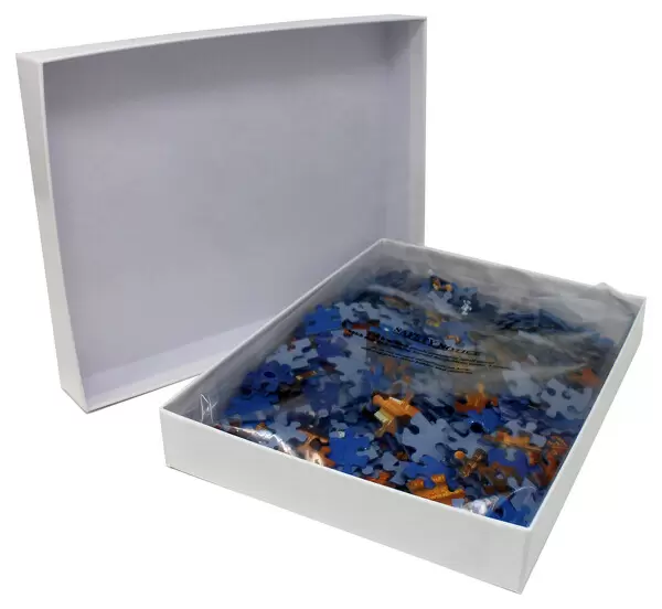
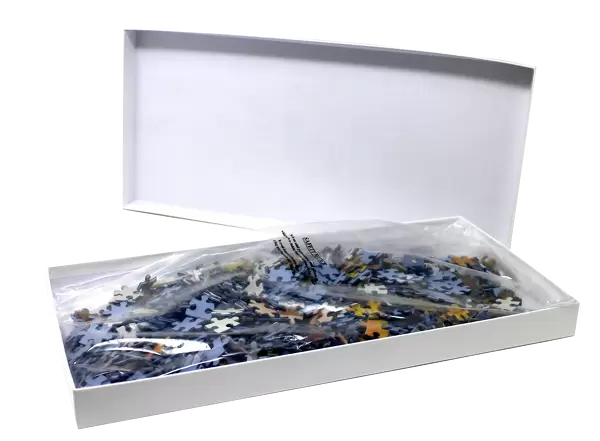
Jigsaw Puzzle (500 Pieces)
Discover the intricacies of human anatomy with Media Storehouse's educational jigsaw puzzles. Our latest addition to the collection is a captivating challenge based on the X-ray image "Osteoarthritis of the Knee, F008 / 3461" by Science Photo Library. This puzzle provides an engaging way to explore the complexities of osteoarthritis, a common degenerative joint disease. Assemble the puzzle pieces to reveal the striking colors and details of this coloured X-ray image, enhancing your understanding of medical anatomy and imaging techniques. Perfect for healthcare professionals, students, or anyone with an interest in the human body.
500 piece puzzles are custom made in the UK and hand-finished on 100% recycled 1.5 mm millboard. There is a level of repetition in jigsaw shapes with each matching piece away from its pair. The completed puzzle measures 38x50cm and is delivered packaged in an attractive presentation box specially designed to fit most letter box slots
Jigsaw Puzzles are an ideal gift for any occasion
Estimated Product Size is 38cm x 50.2cm (15" x 19.8")
These are individually made so all sizes are approximate
Artwork printed orientated as per the preview above, with landscape (horizontal) or portrait (vertical) orientation to match the source image.
EDITORS COMMENTS
This print from Science Photo Library showcases the intricate details of an X-ray image depicting osteoarthritis of the knee. Against a pristine white background, this coloured X-ray offers a glimpse into the complexities of our skeletal system and the impact of illness on our bodies. The image highlights the joint affected by osteoarthritis, revealing signs of discomfort and disorder within it. The abnormality is evident in the bones comprising the knee - femur, tibia, and fibula - which are clearly visible in this side view radiography. This medical illustration serves as a powerful reminder of both the fragility and resilience of our human anatomy. With its scientific imaging quality, this print not only provides valuable insights for healthcare professionals but also sparks curiosity among those interested in medicine. It invites viewers to ponder upon various aspects related to osteoarthritis such as diagnosis, treatment options, and overall patient care. As we gaze at this mesmerizing visual representation captured by SCIENCE PHOTO LIBRARY, we are reminded that despite its unhealthy appearance, there is hope for individuals suffering from osteoarthritis. Through advancements in medical research and compassionate healthcare practices, we strive towards alleviating discomfort caused by such conditions while promoting healthier lives for all.
MADE IN THE UK
Safe Shipping with 30 Day Money Back Guarantee
FREE PERSONALISATION*
We are proud to offer a range of customisation features including Personalised Captions, Color Filters and Picture Zoom Tools
SECURE PAYMENTS
We happily accept a wide range of payment options so you can pay for the things you need in the way that is most convenient for you
* Options may vary by product and licensing agreement. Zoomed Pictures can be adjusted in the Basket.