Antique Framed Print : Osteoarthritis of the knee, X-ray F008 / 3461


![]()
Framed Photos from Science Photo Library
Osteoarthritis of the knee, X-ray F008 / 3461
Osteoarthritis of the knee, coloured X-ray
Science Photo Library features Science and Medical images including photos and illustrations
Media ID 9305141
© SCIENCE PHOTO LIBRARY
Discomfort Femur Fibula Illness Joint Knee Osteoarthritis Radiography Scientific Imaging Tibia Xray Abnormal Disorder Human Skeleton Unhealthy
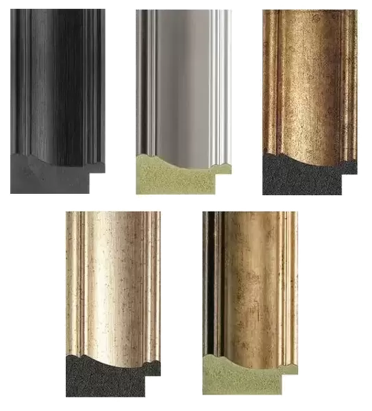
14"x12" (36x31cm) Antique Frame
Bevelled wood effect frame, card mounted, 10x8 archival quality photo print. Overall outside dimensions 14x12 inches (36x31cm). Environmentally and ozone friendly, the Polycore® moulding has the look of real wood, is durable and light and easy to hang. Biodegradable and made with non-chlorinated gases (no toxic fumes) it is efficient; producing 100 tons of polystyrene can save 300 tons of trees! Prints are glazed with lightweight, shatterproof, optical clarity acrylic (providing the same general protection from the environment as glass). The back is stapled hardboard with a sawtooth hanger attached. Note: To minimise original artwork cropping, for optimum layout, and to ensure print is secure, the visible print may be marginally smaller
Bevelled Wood Effect Framed and Mounted Prints - Professionally Made and Ready to Hang
Estimated Image Size (if not cropped) is 20.1cm x 24.4cm (7.9" x 9.6")
Estimated Product Size is 31.2cm x 36.3cm (12.3" x 14.3")
These are individually made so all sizes are approximate
Artwork printed orientated as per the preview above, with portrait (vertical) orientation to match the source image.
EDITORS COMMENTS
This print from Science Photo Library showcases the intricate details of an X-ray image depicting osteoarthritis of the knee. Against a pristine white background, this coloured X-ray offers a glimpse into the complexities of our skeletal system and the impact of illness on our bodies. The image highlights the joint affected by osteoarthritis, revealing signs of discomfort and disorder within it. The abnormality is evident in the bones comprising the knee - femur, tibia, and fibula - which are clearly visible in this side view radiography. This medical illustration serves as a powerful reminder of both the fragility and resilience of our human anatomy. With its scientific imaging quality, this print not only provides valuable insights for healthcare professionals but also sparks curiosity among those interested in medicine. It invites viewers to ponder upon various aspects related to osteoarthritis such as diagnosis, treatment options, and overall patient care. As we gaze at this mesmerizing visual representation captured by SCIENCE PHOTO LIBRARY, we are reminded that despite its unhealthy appearance, there is hope for individuals suffering from osteoarthritis. Through advancements in medical research and compassionate healthcare practices, we strive towards alleviating discomfort caused by such conditions while promoting healthier lives for all.
MADE IN THE UK
Safe Shipping with 30 Day Money Back Guarantee
FREE PERSONALISATION*
We are proud to offer a range of customisation features including Personalised Captions, Color Filters and Picture Zoom Tools
SECURE PAYMENTS
We happily accept a wide range of payment options so you can pay for the things you need in the way that is most convenient for you
* Options may vary by product and licensing agreement. Zoomed Pictures can be adjusted in the Basket.