
Jigsaw Puzzle : False col SEM of skin from blister on human hand


![]()
Jigsaw Puzzles from Science Photo Library
False col SEM of skin from blister on human hand
False colour scanning electron micrograph of human skin from a blister on the palm of the hand (male). The skin on the palm is neatly arranged in ridges (not seen) with sweat pores (seen) appearing as miniature depressions spiralling into the ridge- peaks. The external surface of skin, the epidermis, consists of keratinised, flattened layers of cells. Keratinization occurs when deposits of the fibrous protein keratin are layed down in the cells, causing them to toughen. This outer layer of cells is shed continuously (flaky) & is replaced by progressive movement & maturation of cells from the skin beneath. Magnification: X130 at 35mm size. Original is BW print P710/154
Science Photo Library features Science and Medical images including photos and illustrations
Media ID 6453709
© DR JEREMY BURGESS/SCIENCE PHOTO LIBRARY
Blister Epidermis Skin Surface Sweat Pore
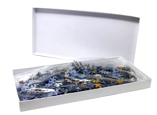
Jigsaw Puzzle (400 Pieces)
Explore the wonders of science with Media Storehouse's Jigsaw Puzzles. Our latest addition to the collection is this captivating False Colour Scanning Electron Micrograph of human skin from a blister on the palm of a hand, courtesy of Science Photo Library. Delve deep into the intricacies of the human body as you piece together this intriguing puzzle, revealing the neatly arranged ridges and visible sweat pores. A perfect blend of education and entertainment, this puzzle is not only a fun activity but also an excellent way to expand your knowledge of the world around us.
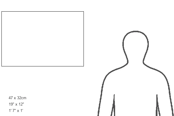
400 piece puzzles are custom made in the UK and hand-finished on 100% recycled 1.5 mm millboard. There is a level of repetition in jigsaw shapes with each matching piece away from its pair. The completed puzzle measures 31x47cm and is delivered packaged in an attractive presentation box specially designed to fit most letter box slots
Jigsaw Puzzles are an ideal gift for any occasion
Estimated Product Size is 47.2cm x 31.5cm (18.6" x 12.4")
These are individually made so all sizes are approximate
Artwork printed orientated as per the preview above, with landscape (horizontal) or portrait (vertical) orientation to match the source image.
EDITORS COMMENTS
This false color scanning electron micrograph showcases a close-up view of human skin from a blister on the palm of a male hand. The intricate ridges, which are not visible in this image, neatly arrange the skin's surface. Delving deeper into the photograph, one can observe miniature depressions spiraling into these ridge-peaks - these are sweat pores. The outer layer of our skin, known as the epidermis, is composed of flattened layers of keratinized cells. Keratinization occurs when deposits of the fibrous protein keratin toughen and fortify these cells. As part of its natural process, this outer layer continuously sheds (hence its flaky appearance) and is replaced by new cells maturing from beneath. At a magnification level of X130 with a 35mm size print, this black-and-white image offers an extraordinary glimpse into the anatomy and structure that make up our largest organ - our skin. It serves as a reminder that even at such microscopic scales, there exists incredible complexity within our bodies.
MADE IN THE UK
Safe Shipping with 30 Day Money Back Guarantee
FREE PERSONALISATION*
We are proud to offer a range of customisation features including Personalised Captions, Color Filters and Picture Zoom Tools
SECURE PAYMENTS
We happily accept a wide range of payment options so you can pay for the things you need in the way that is most convenient for you
* Options may vary by product and licensing agreement. Zoomed Pictures can be adjusted in the Basket.