
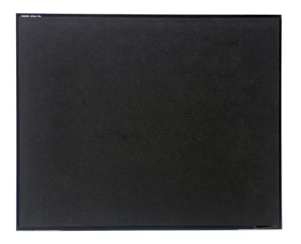
Glass Place Mat : False col SEM of skin from blister on human hand


![]()
Home Decor from Science Photo Library
False col SEM of skin from blister on human hand
False colour scanning electron micrograph of human skin from a blister on the palm of the hand (male). The skin on the palm is neatly arranged in ridges (not seen) with sweat pores (seen) appearing as miniature depressions spiralling into the ridge- peaks. The external surface of skin, the epidermis, consists of keratinised, flattened layers of cells. Keratinization occurs when deposits of the fibrous protein keratin are layed down in the cells, causing them to toughen. This outer layer of cells is shed continuously (flaky) & is replaced by progressive movement & maturation of cells from the skin beneath. Magnification: X130 at 35mm size. Original is BW print P710/154
Science Photo Library features Science and Medical images including photos and illustrations
Media ID 6453709
© DR JEREMY BURGESS/SCIENCE PHOTO LIBRARY
Blister Epidermis Skin Surface Sweat Pore
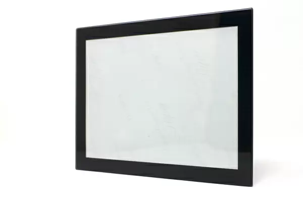
Glass Place Mat (Set of 4)
Set of 4 Glass Place Mats. Stylish and elegant polished safety glass, toughened and heat resistant (275x225mm, 7mm thick). Matching Coasters also available.
Set of 4 Glass Place Mats. Elegant polished safety glass and heat resistant. Matching Coasters may also be available
Estimated Image Size (if not cropped) is 25.4cm x 17.9cm (10" x 7")
Estimated Product Size is 27.5cm x 22.5cm (10.8" x 8.9")
These are individually made so all sizes are approximate
EDITORS COMMENTS
This false color scanning electron micrograph showcases a close-up view of human skin from a blister on the palm of a male hand. The intricate ridges, which are not visible in this image, neatly arrange the skin's surface. Delving deeper into the photograph, one can observe miniature depressions spiraling into these ridge-peaks - these are sweat pores. The outer layer of our skin, known as the epidermis, is composed of flattened layers of keratinized cells. Keratinization occurs when deposits of the fibrous protein keratin toughen and fortify these cells. As part of its natural process, this outer layer continuously sheds (hence its flaky appearance) and is replaced by new cells maturing from beneath. At a magnification level of X130 with a 35mm size print, this black-and-white image offers an extraordinary glimpse into the anatomy and structure that make up our largest organ - our skin. It serves as a reminder that even at such microscopic scales, there exists incredible complexity within our bodies.
MADE IN THE UK
Safe Shipping with 30 Day Money Back Guarantee
FREE PERSONALISATION*
We are proud to offer a range of customisation features including Personalised Captions, Color Filters and Picture Zoom Tools
SECURE PAYMENTS
We happily accept a wide range of payment options so you can pay for the things you need in the way that is most convenient for you
* Options may vary by product and licensing agreement. Zoomed Pictures can be adjusted in the Basket.