Greetings Card : LM of a cross section of cerebellar tissue


![]()
Cards from Science Photo Library
LM of a cross section of cerebellar tissue
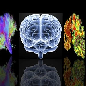
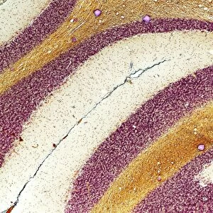
Cerebellar tissue. Light micrograph showing the histological arrangement of a portion of the brain called the cerebellum. The cerebellar cortex (red-orange) is divided into two layers known as the molecular (outer) and granular layers. The junction between these layers is marked by a continuous line of black dots which represent the bodies of the nerve cells known as Purkinje cells. The central core of the cerebellum (light brown) is formed by white matter which consists of densely packed nerve fibres. The cerebellum is part of the central nervous system and is concerned with the maintenance of balance and posture. Magnification: x5 at 10x12cm size
Science Photo Library features Science and Medical images including photos and illustrations
Media ID 6421512
© PASIEKA/SCIENCE PHOTO LIBRARY
Central Nervous System Cerebellum Granular Layer Molecular Layer Purkinje Cell White Matter Brain Light Micrograph
Greetings Card (A5)
Bring a unique touch to your greetings with Media Storehouse's Science-themed card collection. This captivating design features a light micrograph image of cerebellar tissue from Science Photo Library. Delve into the intricacies of the brain with a cross-section view of the cerebellum, showcasing its distinctive red-orange cerebellar cortex. Surprise and delight your recipient with this thoughtful and thought-provoking card, perfect for science enthusiasts, students, or anyone with an appreciation for the wonders of the human body.
Create your own greetings card. Size when folded is A5 (14.5x21cm or 5.7x8.3 inches)
Greetings Cards suitable for Birthdays, Weddings, Anniversaries, Graduations, Thank You and much more
Estimated Image Size (if not cropped) is 14.5cm x 21cm (5.7" x 8.3")
Estimated Product Size is 29cm x 21cm (11.4" x 8.3")
These are individually made so all sizes are approximate
Artwork printed orientated as per the preview above, with portrait (vertical) orientation to match the source image.
EDITORS COMMENTS
This print from Science Photo Library provides a detailed glimpse into the intricate structure of cerebellar tissue. The image showcases a cross section of the brain's cerebellum, highlighting its histological arrangement. The vibrant red-orange color represents the cerebellar cortex, which is divided into two distinct layers: the outer molecular layer and the inner granular layer. A striking feature in this micrograph is the continuous line of black dots that demarcate the junction between these layers. These dots are actually Purkinje cells, nerve cells responsible for transmitting information within the brain. Their presence signifies an important boundary within this complex organ. The light brown central core visible in this image consists of white matter, composed of densely packed nerve fibers. This essential component plays a crucial role in transmitting signals throughout the nervous system. As part of our central nervous system, the cerebellum serves a vital function in maintaining balance and posture. Its intricate architecture can be appreciated through this magnified view captured at 10x12cm size with a magnification factor of x5. This fascinating photograph offers valuable insights into human anatomy, specifically focusing on key elements such as purkinje cells, cortex layers, and white matter within cerebellar tissue. It serves as a testament to Science Photo Library's commitment to providing high-quality scientific imagery for educational purposes.
MADE IN THE UK
Safe Shipping with 30 Day Money Back Guarantee
FREE PERSONALISATION*
We are proud to offer a range of customisation features including Personalised Captions, Color Filters and Picture Zoom Tools
SECURE PAYMENTS
We happily accept a wide range of payment options so you can pay for the things you need in the way that is most convenient for you
* Options may vary by product and licensing agreement. Zoomed Pictures can be adjusted in the Basket.