Greetings Card > Europe > United Kingdom > England > London > Sights > London Eye
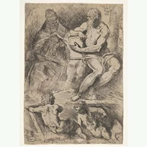
Greetings Card : Facial nerves


![]()
Cards from Science Photo Library
Facial nerves
Facial nerves. Historical anatomical artwork of a side view of a dissected human head showing the nerves (white) and muscles (red) of the face and neck. The facial nerves originate from a nerve called the seventh cranial nerve, also called the facial nerve. The left-hand facial nerve is seen here, emerging from below the ear. It branches out to ennervate the muscles of the face and scalp, and also the salivary glands and lacrimal glands of the eye (for tears). Sensory information comes from the tongue. Separate nerves in the neck run to the back of the scalp and to neck muscles. Artwork from The Nerves of the Human Body (Ed. Jones Quain, London, 1839)
Science Photo Library features Science and Medical images including photos and illustrations
Media ID 6419584
© SHEILA TERRY/SCIENCE PHOTO LIBRARY
1839 Book Dissected Dissection Drawing Face Facial Nerve Jones Quain Lateral Muscles Nerve Nerves People Person Persons Peripheral Profile Side Text Book Nervous System Neurological Neurology Section Sectioned
Greetings Card Large (A4)
Discover the beauty of science with our unique Media Storehouse Greetings Cards. This captivating design features a historical anatomical artwork of a dissected human head, showcasing the intricate details of the Facial Nerves. Explore the complex network of white nerves and red muscles, a testament to the marvels of the human body. Perfect for the science enthusiast or anyone who appreciates the wonders of anatomy, these cards are sure to leave a lasting impression.
Create your own large greetings card. Size when folded is A4 (21x30cm or 8.3x11.7 inches)
Greetings Cards suitable for Birthdays, Weddings, Anniversaries, Graduations, Thank You and much more
Estimated Image Size (if not cropped) is 21cm x 29.7cm (8.3" x 11.7")
Estimated Product Size is 42cm x 29.7cm (16.5" x 11.7")
These are individually made so all sizes are approximate
Artwork printed orientated as per the preview above, with portrait (vertical) orientation to match the source image.
FEATURES IN THESE COLLECTIONS
> Europe
> United Kingdom
> England
> London
> Sights
> London Eye
EDITORS COMMENTS
This 19th-century artwork, titled "Facial Nerves" offers a remarkable glimpse into the intricate anatomy of the human head. A side view of a dissected human head reveals an elaborate network of nerves (depicted in white) and muscles (highlighted in red), showcasing the complexity and beauty of our facial structure. The focus here lies on the seventh cranial nerve, commonly known as the facial nerve, which originates from below the ear. The left-hand facial nerve takes center stage as it branches out to innervate various crucial components of our face and neck. Notably, it supplies motor function to the muscles responsible for facial expressions and scalp movement while also reaching out to salivary glands and lacrimal glands that produce tears for our eyes. Beyond its motor functions, this extraordinary nerve also carries sensory information from our tongue. Additionally, separate nerves extend into the neck region, connecting with muscles at the back of our scalp and neck. Originally featured in "The Nerves of the Human Body" by Jones Quain in 1839, this historical anatomical illustration serves as both a testament to medical knowledge during that era and an artistic masterpiece. Its meticulous details provide valuable insights into neurology and highlight how far we have come in understanding our own bodies throughout history.
MADE IN THE UK
Safe Shipping with 30 Day Money Back Guarantee
FREE PERSONALISATION*
We are proud to offer a range of customisation features including Personalised Captions, Color Filters and Picture Zoom Tools
SECURE PAYMENTS
We happily accept a wide range of payment options so you can pay for the things you need in the way that is most convenient for you
* Options may vary by product and licensing agreement. Zoomed Pictures can be adjusted in the Basket.