Glass Frame : Coloured X-ray of hip fracture due to osteoporosis


![]()
Mounted Prints from Science Photo Library
Coloured X-ray of hip fracture due to osteoporosis
Osteoporosis fracture. Coloured X-ray of the hip of an elderly woman with a fracture (centre left, yellow) caused by osteoporosis (brittle bone disease). The femur (thigh bone, lower right) articulates with the pelvis at the hip socket (centre). Osteoporosis causes a loss in bone density, making the bones thinner, more brittle and more likely to break. In this case the pubis bone of the pelvis was fractured by a fall. Osteoporosis is most common in women after the menopause as their ovaries no longer produce the oestrogen hormones which help to maintain bone mass
Science Photo Library features Science and Medical images including photos and illustrations
Media ID 6414942
© MEDICAL PHOTO NHS LOTHIAN/SCIENCE PHOTO LIBRARY
Brittle Bone Disease Fracture Hip Fracture Osteoporosis Pubis Condition Disorder Elderly Woman Health Care Pelvis
7"x5" Glass Mount
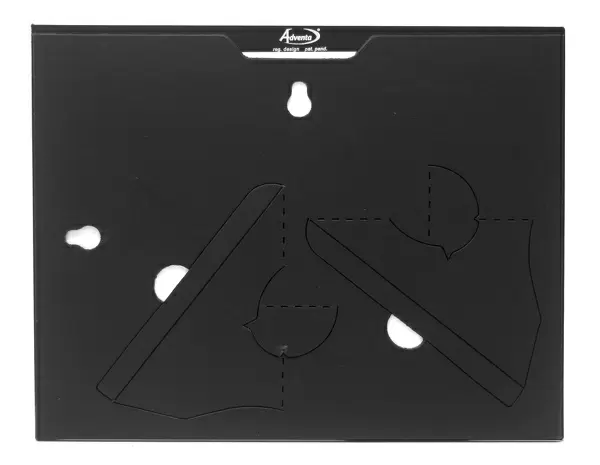
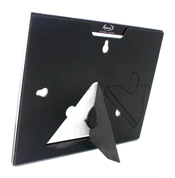
Wall mounted or free-standing, these black edged glass frames feature a smooth chamfered edge and a stylish black border (on back face of the glass). Manufactured from 4mm thick glass, Glass Mounts are a durable, professional way of displaying and protecting your prints. Your 7x5 print is slotted into the back of the frame so can easily be changed if needed.
Tempered Glass Mounts are ideal for wall display, plus the smaller sizes can also be used free-standing via an integral stand
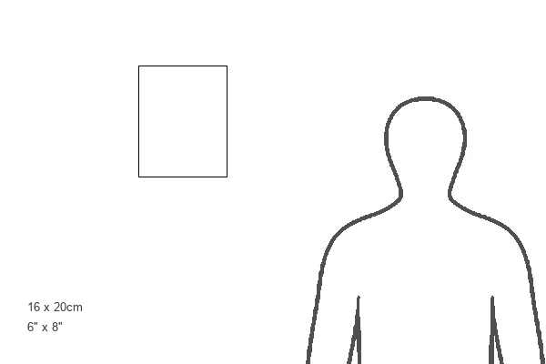
Estimated Image Size (if not cropped) is 12.7cm x 17.7cm (5" x 7")
Estimated Product Size is 16.2cm x 20.3cm (6.4" x 8")
These are individually made so all sizes are approximate
Artwork printed orientated as per the preview above, with portrait (vertical) orientation to match the source image.
EDITORS COMMENTS
This print showcases the devastating effects of osteoporosis on an elderly woman's hip. The vibrant colors highlight a fracture (yellow) caused by this debilitating bone disease, emphasizing the severity of the condition. Osteoporosis, known as brittle bone disease, leads to a loss in bone density and makes bones thinner and more susceptible to fractures. In this particular case, a fall resulted in a fractured pubis bone of the pelvis. The intricate X-ray reveals how the femur articulates with the hip socket at its center-right position. Osteoporosis primarily affects women after menopause due to decreased production of estrogen hormones that play a crucial role in maintaining bone mass. The image serves as a powerful reminder of the importance of early detection and prevention strategies for osteoporosis. It highlights both the fragility and resilience of our skeletal system while underscoring the need for proper medical care and attention to mitigate such fractures. Science Photo Library has expertly captured this moment, shedding light on one aspect of osteoporosis through their remarkable photography skills. This thought-provoking image not only educates viewers about this prevalent health issue but also emphasizes society's responsibility towards promoting better healthcare practices for all individuals affected by osteoporosis.
MADE IN THE UK
Safe Shipping with 30 Day Money Back Guarantee
FREE PERSONALISATION*
We are proud to offer a range of customisation features including Personalised Captions, Color Filters and Picture Zoom Tools
SECURE PAYMENTS
We happily accept a wide range of payment options so you can pay for the things you need in the way that is most convenient for you
* Options may vary by product and licensing agreement. Zoomed Pictures can be adjusted in the Basket.