
Glass Frame > Animals > Mammals > Muridae > Blue-grey Mouse
Glass Frame : Coloured SEM of capillaries of the gall bladder


![]()
Mounted Prints from Science Photo Library
Coloured SEM of capillaries of the gall bladder
Capillaries of the gall bladder. Coloured Scanning Electron Micrograph (SEM) of blood vessels lining the wall of the gall bladder. Here, a rich network of fine capillaries is seen (pink) branching off a blood vessel (grey). Beneath this network are a number of larger blood vessels (blue). This vascular system occurs in the submucosal layer of the gall bladder. Blood and lymphatic vessels serve to drain water reabsorbed from bile during bile formation. The gall bladder is a muscular sac, attached to the liver, which collects and concentrates bile, a substance which aids in fat digestion. Magnification: x100 at 6x7cm size. Magnification: x125 at 4x5 inch size
Science Photo Library features Science and Medical images including photos and illustrations
Media ID 6420704
© PROFESSOR P.M. MOTTA, A. CAGGIATI & G. MACCHIARELLI/SCIENCE PHOTO LIBRARY
Blood Capillaries Capillary Gall Bladder Vessel Vessels
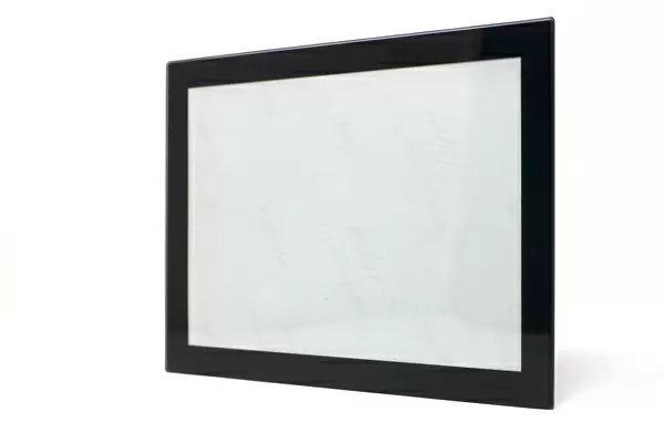
7"x5" Glass Mount
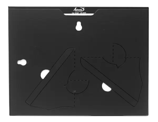
Wall mounted or free-standing, these black edged glass frames feature a smooth chamfered edge and a stylish black border (on back face of the glass). Manufactured from 4mm thick glass, Glass Mounts are a durable, professional way of displaying and protecting your prints. Your 7x5 print is slotted into the back of the frame so can easily be changed if needed.
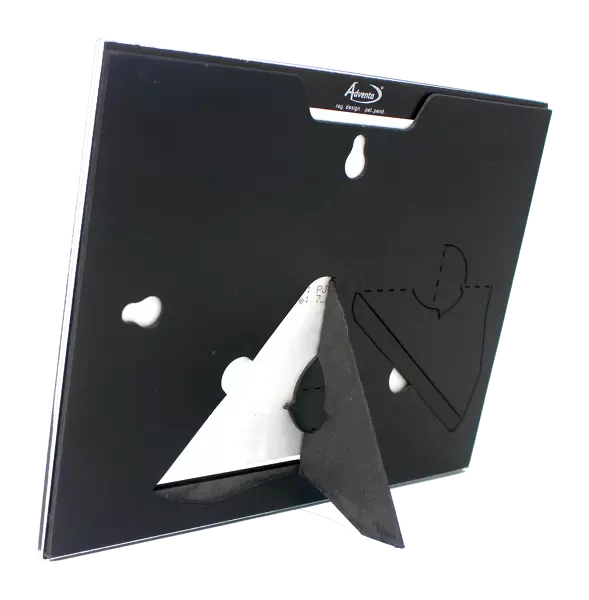
Tempered Glass Mounts are ideal for wall display, plus the smaller sizes can also be used free-standing via an integral stand
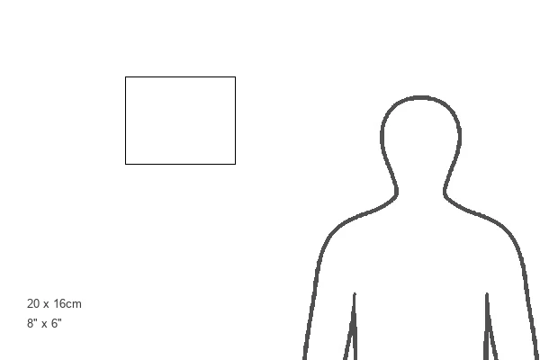
Estimated Image Size (if not cropped) is 17.7cm x 12.7cm (7" x 5")
Estimated Product Size is 20.3cm x 16.2cm (8" x 6.4")
These are individually made so all sizes are approximate
Artwork printed orientated as per the preview above, with landscape (horizontal) orientation to match the source image.
EDITORS COMMENTS
This print showcases the intricate network of capillaries that line the walls of the gall bladder. In this coloured Scanning Electron Micrograph (SEM), we are presented with a mesmerizing view of blood vessels in various shades and sizes. The fine capillaries, depicted in a delicate pink hue, branch off from a larger grey blood vessel, creating an elaborate web-like structure. Beneath this network lies a series of larger blue blood vessels, adding depth and complexity to the vascular system within the submucosal layer of the gall bladder. These vital channels play a crucial role in draining water reabsorbed from bile during its formation process. The gall bladder itself is a muscular sac attached to the liver, responsible for collecting and concentrating bile – an essential substance aiding in fat digestion. This remarkable image offers us an up-close look at one aspect of this organ's intricate inner workings. With magnifications reaching x100 at 6x7cm size and x125 at 4x5 inch size, every minute detail is brought to life through this photograph by Science Photo Library. It serves as both an educational tool for understanding human anatomy and as a testament to nature's awe-inspiring design found within our own bodies.
MADE IN THE UK
Safe Shipping with 30 Day Money Back Guarantee
FREE PERSONALISATION*
We are proud to offer a range of customisation features including Personalised Captions, Color Filters and Picture Zoom Tools
SECURE PAYMENTS
We happily accept a wide range of payment options so you can pay for the things you need in the way that is most convenient for you
* Options may vary by product and licensing agreement. Zoomed Pictures can be adjusted in the Basket.