Glass Coaster : LM of human embryo at two-cell stage


![]()
Home Decor from Science Photo Library
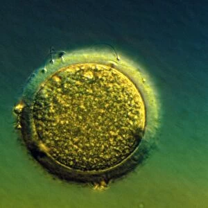
LM of human embryo at two-cell stage
Light micrograph of a human zygote (the primitive embryo), composed of two cells following its first mitotic division - the initial transformation from a single cell organism to one composed of millions of cells. The two cells (called blastomeres) both undergo similar cleavage; successive divisions result in the formation of a hollow ball of cells (the blastocyst) with a localised thickening (the inner cell mass) that will develop into the actual embryo. Development of the blastocyst occurs before it implants into the uterine wall. The site of implantation determines the subsequent position of the placenta
Science Photo Library features Science and Medical images including photos and illustrations
Media ID 6454823
© ANDY WALKER, MIDLAND FERTILITY SERVICES/ SCIENCE PHOTO LIBRARY
Blastomere Embryo Light Micrograph
Glass Coaster
Individual Glass Coaster. Stylish and elegant polished safety glass, toughened and heat resistant (10x10cm, 7mm thick). Price shown is per individual coaster.
Individual Glass Coaster. Elegant polished safety toughened glass and heat resistant, matching Place Mats are also available
Estimated Image Size (if not cropped) is 7.6cm x 5.1cm (3" x 2")
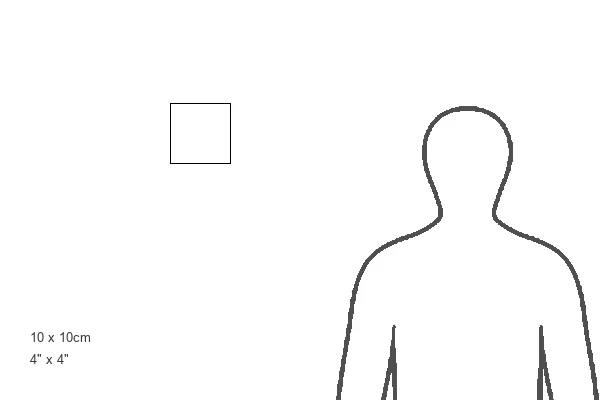
Estimated Product Size is 10cm x 10cm (3.9" x 3.9")
These are individually made so all sizes are approximate
Artwork printed orientated as per the preview above, with landscape (horizontal) orientation to match the source image.
EDITORS COMMENTS
This print captures the mesmerizing beauty of a human zygote at its two-cell stage. The delicate and intricate details showcased in this light micrograph highlight the miraculous transformation from a single cell organism to a complex structure composed of millions of cells. At this early stage, known as the blastomere phase, both cells undergo similar cleavage, dividing successively to form a hollow ball of cells called the blastocyst. Within this spherical arrangement, there is a localized thickening known as the inner cell mass that will eventually develop into the actual embryo. The development of the blastocyst occurs prior to its implantation into the uterine wall. This critical process determines not only where it attaches but also influences the subsequent position of the placenta. Through this remarkable image, we gain insight into one of nature's most awe-inspiring phenomena – embryonic development. It serves as a reminder of our own origin and underscores how every individual begins their journey with such incredible complexity and potential for growth. Science Photo Library has once again captured an extraordinary moment in scientific exploration by providing us with this striking visual representation. Whether you are fascinated by biology or simply appreciate artistry in science, this print offers an opportunity for contemplation and wonderment at life's earliest stages.
MADE IN THE UK
Safe Shipping with 30 Day Money Back Guarantee
FREE PERSONALISATION*
We are proud to offer a range of customisation features including Personalised Captions, Color Filters and Picture Zoom Tools
SECURE PAYMENTS
We happily accept a wide range of payment options so you can pay for the things you need in the way that is most convenient for you
* Options may vary by product and licensing agreement. Zoomed Pictures can be adjusted in the Basket.