Fraud Blocker Skip to main content

Vascular Collection (page 8)

"Exploring the Intricate World Anatomy: From Brain Blood Vessels to Maize Roots" Delve into the fascinating realm anatomy

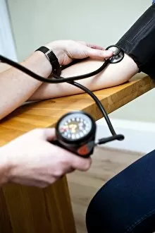
Background imageVascular Collection: Blood pressure measurement C013 / 7569

Blood pressure measurement C013 / 7569
Blood pressure measurement. Patient having their blood pressure taken at a doctors surgery

Background imageVascular Collection: Blood pressure measurement C013 / 7568

Blood pressure measurement C013 / 7568
Blood pressure measurement. Patient having their blood pressure taken at a doctors surgery

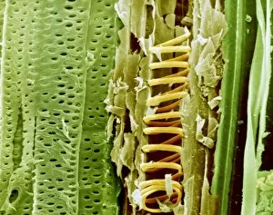
Background imageVascular Collection: Buttercup stem, SEM

Buttercup stem, SEM
Buttercup stem. Coloured scanning electron micrograph (SEM) of a section through the stem of a buttercup (Ranunculus sp.) plant

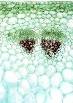
Background imageVascular Collection: Groundsel stem, light micrograph

Groundsel stem, light micrograph
Groundsel stem. Light micrograph of a section through a groundsel stem (Senecio vulgaris), showing xylem vessels (brown, upper centre)

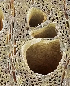
Background imageVascular Collection: Elm wood, SEM

Elm wood, SEM
Elm wood. Coloured scanning electron micrograph (SEM) of a transverse section through wood from an elm tree (Ulmus procera). Xylem vessels (holes) are seen in the wood

Background imageVascular Collection: Silurian landplants

Silurian landplants
An artists impression of the the earliest vascular plants which developed beside freshwater pools during the Silurian (443 to 417 million years ago)

Background imageVascular Collection: Tomato (Solanum sp. ) Green Zebra, close-up of fruit, with Fusarium Wilt

Tomato (Solanum sp. ) Green Zebra, close-up of fruit, with Fusarium Wilt (Fusarium oxysporum lycopersici)
Tomato (Solanum sp.) Green Zebra, close-up of fruit, with Fusarium Wilt (Fusarium oxysporum lycopersici) causes vascular wilt of leaves, U.S.A

Background imageVascular Collection: Giant Horsetail (Equisetum telmateia) patch growing on pebbles at edge of beach, Bembridge

Giant Horsetail (Equisetum telmateia) patch growing on pebbles at edge of beach, Bembridge, Isle of Wight, England, june

Background imageVascular Collection: Field Horsetail (Equisetum arvense) close-up of fertile cones, Sheffield, South Yorkshire

Field Horsetail (Equisetum arvense) close-up of fertile cones, Sheffield, South Yorkshire, England, may

Background imageVascular Collection: Blood vessels in a frog ovary, SEM

Blood vessels in a frog ovary, SEM
Blood vessels in the ovary of a female frog, coloured scanning electron micrograph (SEM). Frogs produce hundreds of eggs, which are expelled and fertilised in the water

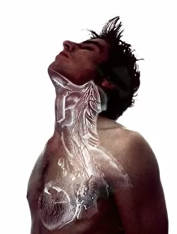
Background imageVascular Collection: Upper body anatomy

Upper body anatomy
MODEL RELEASED. Upper body anatomy. Mans upper body overlaid with an artwork of the heart and the blood vessels and musculature of the chest and neck

Background imageVascular Collection: Female body anatomy

Female body anatomy, artwork

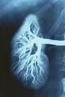
Background imageVascular Collection: Renal blood vessels, X-ray

Renal blood vessels, X-ray
Renal blood vessels. Angiogram (X-ray) of blood vessels in a healthy kidney. The large artery entering the kidney is the renal artery, which branches into many smaller capillaries inside the kidney

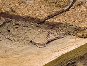
Background imageVascular Collection: Outer layers of the eye, SEM

Outer layers of the eye, SEM
Outer layers of the eye. Coloured scanning electron micrograph (SEM) of the choroid (top) and sclera (bottom) layers of the eye. The sclera is the white outer layer of the eye

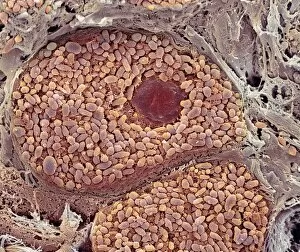
Background imageVascular Collection: Choroid layer of the eye, SEM

Choroid layer of the eye, SEM
Choroid layer of the eye. Coloured scanning electron micrograph (SEM) of a section through the choroid layer of the eye. A pigment cell (dark brown) is at upper centre

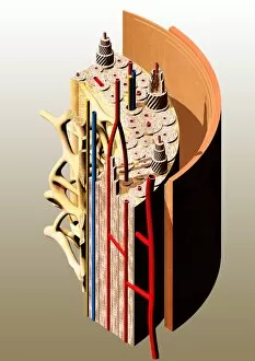
Background imageVascular Collection: Bone anatomy

Bone anatomy. Computer artwork showing the anatomy of a normal bone shaft. An outer layer of compact bone is made up of cylindrical units (osteons, black stripes)

Background imageVascular Collection: Bone structural unit

Bone structural unit. Computer artwork of an osteon, the basic structural unit of compact bone. Compact bone is the outer layer of a bone shaft, and contains numerous osteons

Background imageVascular Collection: Blood vessel, SEM

Blood vessel, SEM
Blood vessel. Coloured scanning electron micrograph (SEM) of a congested blood vessel in human foreskin. The vessel is filled with erythrocytes (red blood cells)

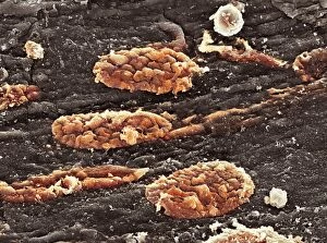
Background imageVascular Collection: Blood clots, SEM

Blood clots, SEM
Blood clots. Coloured scanning electron micrograph (SEM) of small blood clots (red) in a blood vessel. The clots consist of red blood cells, white blood cells, and platelets

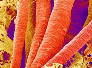
Background imageVascular Collection: Brain arteries, SEM

Brain arteries, SEM
Brain arteries. Coloured scanning electron micrograph (SEM) of arteries from the brain. Arteries carry oxygenated blood from the heart around the body

Background imageVascular Collection: Brain blood vessels, SEM

Brain blood vessels, SEM
Brain blood vessels. Coloured scanning electron micrograph (SEM) of blood vessels from the brain. Veins, which carry deoxygenated blood towards the heart, are yellow and arteries

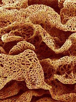
Background imageVascular Collection: Blood vessels from intestinal wall, SEM

Blood vessels from intestinal wall, SEM
Blood vessels. Coloured scanning electron micrograph (SEM) of blood vessels from villi in the duodenum, part of the small intestine

Background imageVascular Collection: Sickle cell anaemia

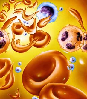
Sickle cell anaemia. Artwork of red blood cells in sickle cell anaemia (drepanocytosis). The sickle- shaped red blood cells (some seen at upper left) contrast with normal red blood cells (rounded)

Background imageVascular Collection: Medical nanorobots on red blood cell

Medical nanorobots on red blood cell
Medical nanorobots. Computer artwork of medical nanorobots on red blood cells inside a human body. Microscopic robot technology could be developed in the future to treat diseases in new ways

Background imageVascular Collection: Angioplasty techniques

Angioplasty techniques. Computer illustration of balloon and laser angioplasty. Angioplasty is a surgical procedure to unblock


All Professionally Made to Order for Quick Shipping

"Exploring the Intricate World Anatomy: From Brain Blood Vessels to Maize Roots" Delve into the fascinating realm anatomy, where a complex network of blood vessels ensures our bodies function seamlessly. Witness the marvels of this intricate system through various historical artworks and scientific imagery. Travel back in time to 1981 with a mesmerizing 3D angiogram C007, showcasing the intricate web of brain blood vessels. Marvel at how these delicate pathways nourish our most vital organ, ensuring its optimal performance. Step into the world of cardiovascular system exploration as you uncover historical artwork depicting its complexity. Admire exquisite neck anatomy illustrations from the 19th century, capturing every detail with artistic precision. Discover the hidden beauty within human heart anatomy through stunning artwork that showcases its awe-inspiring structure. Dive deep into cell structures that make up this remarkable organ, unraveling its secrets one layer at a time. Journey through time as you explore arterial systems depicted in captivating 18th-century artwork. Be captivated by their elegance and understand their crucial role in maintaining overall health and vitality. Immerse yourself in neck vascular anatomy captured by historical artists who sought to document this enigmatic region's intricacies. Let your eyes trace each artery's path as it weaves through muscles and tissues. Witness X-ray images revealing neck and shoulder arteries' hidden mysteries—an opportunity to glimpse beneath our skin's surface and appreciate nature's design flawlessly crafted for optimum functionality. Marvel at Salvia officinalis or Common Sage plant—a botanical wonder known for promoting healthy circulation—its vibrant leaves symbolizing lifeblood coursing through our veins like an eternal river. Transport yourself back to 1825 when male groin arteries were meticulously illustrated—an homage to medical pioneers who unraveled body secrets long before modern technology existed. Uncover artistry behind blood coagulation cascade depicted in breathtaking artwork C016 / 9873.