Fraud Blocker Skip to main content

Vascular Collection (page 2)

"Exploring the Intricate World Anatomy: From Brain Blood Vessels to Maize Roots" Delve into the fascinating realm anatomy

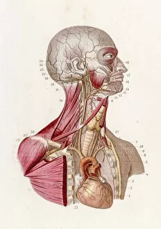
Background imageVascular Collection: Vascular system anatomy engraving 1886

Vascular system anatomy engraving 1886
A Text book of Naked Eye Anatomy

Background imageVascular Collection: Field of European white water lilies (Nymphaea alba) in lake with trees on shore

Field of European white water lilies (Nymphaea alba) in lake with trees on shore and morning haze, Noordhollands Duinreservaat, Noord-Holland, the Netherlands

Background imageVascular Collection: Aloe, flowering Aloe vera plant

Aloe, flowering Aloe vera plant

Background imageVascular Collection: Stroke, MRI and 3D CT scans C016 / 6419

Stroke, MRI and 3D CT scans C016 / 6419
Stroke. Coloured composite image of a magnetic resonance imaging (MRI) scan of the brain and 2D and 3D computed tomography (CT)

Background imageVascular Collection: Female skeleton and organs, artwork

Female skeleton and organs, artwork
Female skeleton and organs and blood vessels, artwork

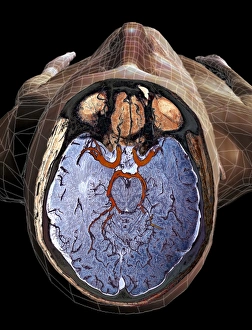
Background imageVascular Collection: Brain blood vessels, artwork

Brain blood vessels, artwork
Brain blood vessels. Computer artwork showing the blood vessels that supply the human brain. The front of the brain is at left

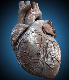
Background imageVascular Collection: Heart of stone

Heart of stone, computer artwork. The heart is a hollow sac (lower frame) of muscular tissue that pumps blood around the body. Small surface coronary blood vessels supply the heart muscle with blood

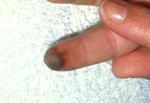
Background imageVascular Collection: Finger of patient with peripheral vasoconstriction

Finger of patient with peripheral vasoconstriction
Peripheral vasoconstriction. Finger showing necrosis or gangrene (tissue death) due to peripheral vasoconstriction. The constriction of the fingers blood vessels means that not enough blood is

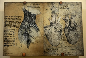
Background imageVascular Collection: Cardiovascular system Leonardo da vincis drawing

Cardiovascular system Leonardo da vincis drawing
Cardiovascular systems drawing. In 1513 Leonardo da Vinci to study the heart and the circulatory system through animal dissections. Windsor, Royal Library 19073, 1510-1513

Background imageVascular Collection: Fern Leaves Touching One Another

Fern Leaves Touching One Another
A fern is a member of a group of about 10, 560 known extant species of vascular plants that reproduce via spores and have neither seeds nor flowers. They differ from mosses by being vascular (i.e)

Background imageVascular Collection: Ischaemia, digital angiogram

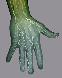
Ischaemia, digital angiogram
image d angiographie numerisee( non soustraite) de la main gauche, en vue de face, par catheterisme hyperselectif des arteres du membre superieur gauche, sous anasthesie generale

Background imageVascular Collection: Neck and head arteries, MRA scan C016 / 6449

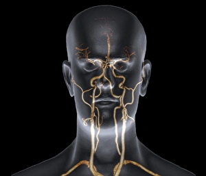
Neck and head arteries, MRA scan C016 / 6449
Neck and head arteries. Coloured magnetic resonance angiography (MRA) scan of a normal carotid system of a 36 year old patient

Background imageVascular Collection: Lung alveoli anatomy, artwork C016 / 7680

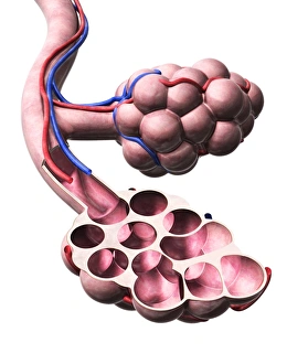
Lung alveoli anatomy, artwork C016 / 7680
Lung alveoli anatomy. Artwork showing the structure of the lung alveoli, where gas exchange occurs between the respiratory and circulatory systems

Background imageVascular Collection: Balloon angioplasty, X-ray

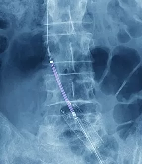
Balloon angioplasty, X-ray
Balloon angioplasty. Coloured X-ray of the abdomen of a 45-year-old patient with a history of thrombosis (blood clots) in the inferior vena cava (vein), which have narrowed (stenosed)

Background imageVascular Collection: Brain blood supply, MRI scan C018 / 0432

Brain blood supply, MRI scan C018 / 0432
Brain haemorrhage. Coloured magnetic resonance imaging (MRI) scan of the brain of a 49 year old patient with a subdural haematoma (red)

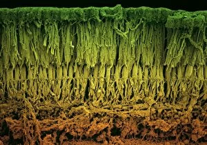
Background imageVascular Collection: Retina layers, SEM C018 / 0520

Retina layers, SEM C018 / 0520
Retina layers, coloured scanning electron micrograph (SEM). The retina of the human eye is found on the inside of the eyeball and is formed from a number of layers

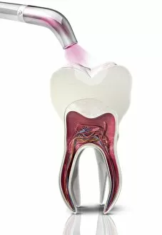
Background imageVascular Collection: Dental filling polymerisation, artwork C014 / 2025

Dental filling polymerisation, artwork C014 / 2025
Dental filling polymerisation. Artwork of a cross-section of a human tooth, with a filling (white) being set using an ultraviolet light lamp tool

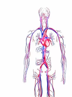
Background imageVascular Collection: Human circulatory system

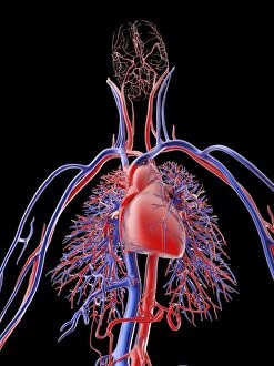
Human circulatory system, computer artwork. Veins (blue) carry deoxygenated blood towards the heart. Arteries (red) carry oxygenated blood from the heart around the body

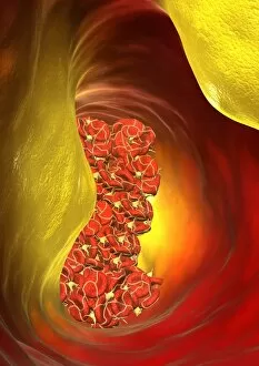
Background imageVascular Collection: Blocked artery, computer artwork

Blocked artery, computer artwork
Blocked artery. Computer artwork of a clot of blood cells (thrombus, red) in an artery affected by atherosclerosis. This is a narrowing of an artery due to fatty deposits (yellow) on its inner walls

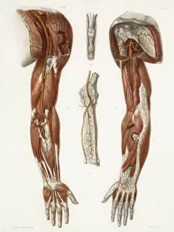
Background imageVascular Collection: Arm anatomy, historical artwork

Arm anatomy, historical artwork. 19th Century hand coloured lithographic print showing the arteries (red), tendons (white) and muscle structure (brown) of the human upper limbs. Fig

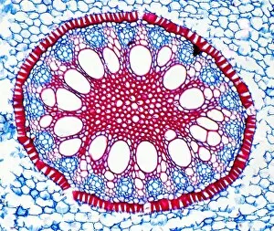
Background imageVascular Collection: Iris root, light micrograph

Iris root, light micrograph
Iris root. Light micrograph of a section through the root of an Iris plant (Iris germanica) showing a vascular cylinder. The cylinder is comprised of a central cluster of parenchyma cells (red)

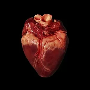
Background imageVascular Collection: Pigs heart

Pigs heart. The heart of a pig is anatomically similar to that of a human. The heart is a hollow muscular organ that pumps blood around the body

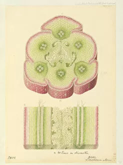
Background imageVascular Collection: Bletia purpurea

Bletia purpurea - Bletia purpurea. Longidunal and traverse section of the ovary. Watercolour by Franz Bauer, 1801. Paper, watercolour. Botany Library, Bauer Unit, Unfinished Drawings of Plants 3: no

Background imageVascular Collection: Wood Horsetail (Equisetum sylvaticum) fertile frond, Powys, Wales, June

Wood Horsetail (Equisetum sylvaticum) fertile frond, Powys, Wales, June

Background imageVascular Collection: Field Horsetail (Equisetum arvense) two fresh shoots, Blashford Lakes Nature Reserve, Avon Valley

Field Horsetail (Equisetum arvense) two fresh shoots, Blashford Lakes Nature Reserve, Avon Valley, New Forest N. P
Field Horsetail (Equisetum arvense) two fresh shoots, Blashford Lakes Nature Reserve, Avon Valley, New Forest N.P. Hampshire, England, March

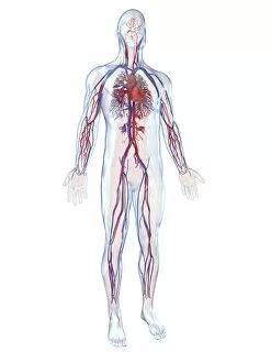
Background imageVascular Collection: Cardiovascular system, artwork F006 / 4620

Cardiovascular system, artwork F006 / 4620
Cardiovascular system, computer artwork

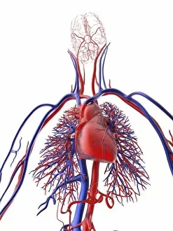
Background imageVascular Collection: Cardiovascular system, artwork F006 / 4611

Cardiovascular system, artwork F006 / 4611
Cardiovascular system, computer artwork

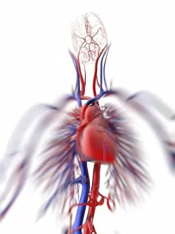
Background imageVascular Collection: Cardiovascular system, artwork F006 / 4600

Cardiovascular system, artwork F006 / 4600
Cardiovascular system, computer artwork

Background imageVascular Collection: Cardiovascular system, artwork F006 / 4596

Cardiovascular system, artwork F006 / 4596
Cardiovascular system, computer artwork

Background imageVascular Collection: Cardiovascular system, artwork F006 / 4602

Cardiovascular system, artwork F006 / 4602
Cardiovascular system, computer artwork

Background imageVascular Collection: Tablet computer, heart attack artwork F006 / 4625

Tablet computer, heart attack artwork F006 / 4625
Tablet computer showing conceptual artwork of a heart attack

Background imageVascular Collection: Blood clot, SEM P260 / 0123

Blood clot, SEM P260 / 0123
Blood clot. Coloured scanning electron micrograph (SEM) of a blood clot. The red blood cells (erythrocytes) are trapped in filaments of fibrin protein (pink)

Background imageVascular Collection: Blood vessel interior, artwork

Blood vessel interior, artwork
Blood vessel interior, computer artwork


All Professionally Made to Order for Quick Shipping

"Exploring the Intricate World Anatomy: From Brain Blood Vessels to Maize Roots" Delve into the fascinating realm anatomy, where a complex network of blood vessels ensures our bodies function seamlessly. Witness the marvels of this intricate system through various historical artworks and scientific imagery. Travel back in time to 1981 with a mesmerizing 3D angiogram C007, showcasing the intricate web of brain blood vessels. Marvel at how these delicate pathways nourish our most vital organ, ensuring its optimal performance. Step into the world of cardiovascular system exploration as you uncover historical artwork depicting its complexity. Admire exquisite neck anatomy illustrations from the 19th century, capturing every detail with artistic precision. Discover the hidden beauty within human heart anatomy through stunning artwork that showcases its awe-inspiring structure. Dive deep into cell structures that make up this remarkable organ, unraveling its secrets one layer at a time. Journey through time as you explore arterial systems depicted in captivating 18th-century artwork. Be captivated by their elegance and understand their crucial role in maintaining overall health and vitality. Immerse yourself in neck vascular anatomy captured by historical artists who sought to document this enigmatic region's intricacies. Let your eyes trace each artery's path as it weaves through muscles and tissues. Witness X-ray images revealing neck and shoulder arteries' hidden mysteries—an opportunity to glimpse beneath our skin's surface and appreciate nature's design flawlessly crafted for optimum functionality. Marvel at Salvia officinalis or Common Sage plant—a botanical wonder known for promoting healthy circulation—its vibrant leaves symbolizing lifeblood coursing through our veins like an eternal river. Transport yourself back to 1825 when male groin arteries were meticulously illustrated—an homage to medical pioneers who unraveled body secrets long before modern technology existed. Uncover artistry behind blood coagulation cascade depicted in breathtaking artwork C016 / 9873.