Fraud Blocker Skip to main content

Neuroscience Collection (page 7)

"Unraveling the Mysteries of the Mind: Exploring Neuroscience through Art and Science" Delve into the intricate world of neuroscience

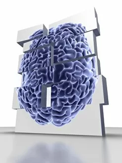
Background imageNeuroscience Collection: Building blocks with brain, artwork

Building blocks with brain, artwork
Conceptual computer artwork of building blocks with the top view of a brain, representing Alzheimers desease or brain disorder

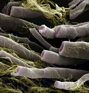
Background imageNeuroscience Collection: Nerve fibres, SEM

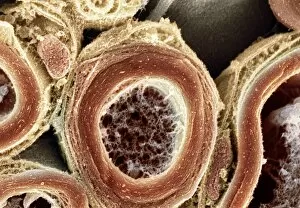
Nerve fibres, SEM
Myelinated nerve fibres, coloured scanning electron micrograph (SEM). The myelin sheath is grey, the axoplasm pink and the endoneurium (connective tissue) yellow

Background imageNeuroscience Collection: Parkinsons disease nerve cells, artwork

Parkinsons disease nerve cells, artwork
Parkinsons disease nerve cells. Computer artwork of human nerve cells (neurons, green) affected by Lewy bodies (orange) in the brain of someone with Parkinsons disease

Background imageNeuroscience Collection: Brain size, conceptual image

Brain size, conceptual image
Brain size. Conceptual image of a human brain wrapped in a tape measure. This image represents the research done on relative brain sizes, which vary between different species

Background imageNeuroscience Collection: Spinal cord, SEM

Spinal cord, SEM
Spinal cord. Coloured scanning electron micrograph (SEM) of a section through a spinal cord, showing the grey matter (green) and white matter (blue)

Background imageNeuroscience Collection: Optic nerve fibres, SEM

Optic nerve fibres, SEM
Optic nerve fibres. Coloured scanning electron micrograph (SEM) of axons from the optic nerve. Connective tissue fibres (thread-like) are also visible

Background imageNeuroscience Collection: Brain lining, SEM

Brain lining, SEM
Brain lining. Coloured scanning electron micrograph (SEM) of the lining of the brain, showing the ciliary hairs (green) of ependymal cells

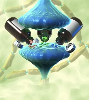
Background imageNeuroscience Collection: Neurological drug treatment, artwork

Neurological drug treatment, artwork
Neurological drug treatment, conceptual image. Computer artwork representing the use of drugs that affect neural networks by altering the transmission of neurotransmitters across synapses (blue)

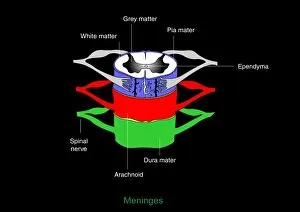
Background imageNeuroscience Collection: Meninges, artwork

Meninges, artwork
Meninges. Computer artwork showing the meninges surrounding the spinal cord. The meninges are the three membranes that envelop the brain and spinal cord

Background imageNeuroscience Collection: Brain anatomy, 3D artwork

Brain anatomy, 3D artwork
Brain anatomy. 3D artwork of a cut-away model of the human brain. At bottom is the brainstem, which controls automatic functions

Background imageNeuroscience Collection: Nerve cell anatomy, artwork

Nerve cell anatomy, artwork
Nerve cell anatomy. Computer artwork showing the organelles in a neurone (nerve cell). At centre is nucleus (grey) and nucleolus (black)

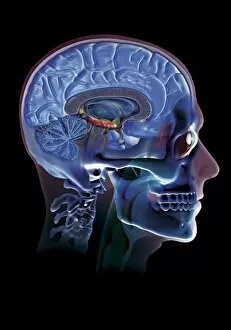
Background imageNeuroscience Collection: Human head, artwork

Human head, artwork
Human head. Computer artwork of a human head showing the skull, cervical (neck) vertebrae and brain inside

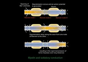
Background imageNeuroscience Collection: Saltatory conduction of nerve impulse

Saltatory conduction of nerve impulse, computer artwork. Nerve impulses move along myelinated nerves by saltatory conduction. The myelin (orange, striped) does not cover the entire axon

Background imageNeuroscience Collection: Synapse, artwork

Synapse, artwork
Synapse. Computer artwork of a synapse, the junction between nerve cells. Synapses transmit electrical signals from one nerve cell to the next

Background imageNeuroscience Collection: Alzheimers disease, computer artwork

Alzheimers disease, computer artwork
3D computer artwork depicting senile amyloid plaque (brown) in the brain, which kill surrounding neurons (blue). Large numbers of senile plaques are characteristic features of Alzheimers disease

Background imageNeuroscience Collection: Bovine prion protein, molecular model

Bovine prion protein, molecular model
Bovine prion protein. Computer model showing the molecular structure of a bovine (cow) prion protein. Prions are abnormal proteins that cause a group of fatal neurodegenerative diseases including BSE

Background imageNeuroscience Collection: Neuromuscular junctions, artwork

Neuromuscular junctions, artwork
Neuromuscular junctions. Computer artwork of junctions between nerves (thread-like objects) and a muscle (purple surface)

Background imageNeuroscience Collection: Internal brain anatomy, artwork

Internal brain anatomy, artwork
Internal brain anatomy. Artwork of a male head and brain from the side, showing the internal brain anatomy. The highly folded area is the cerebrum, which is responsible for conscious thought

Background imageNeuroscience Collection: Synapses, artwork

Synapses, artwork
Synapses. Computer artwork of synapses, the junctions between the ends (green, swollen) of two nerve cells (neurons). Signals are passed along nerve cells in the form of an electrical impulse

Background imageNeuroscience Collection: Brain, artwork

Brain, artwork
Brain. Computer artwork of a side view of a healthy male brain. The front of the brain is at left. The highly folded area is the cerebrum, which is responsible for conscious thought

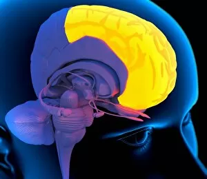
Background imageNeuroscience Collection: Frontal lobe in the brain, artwork

Frontal lobe in the brain, artwork
Frontal lobe in the brain. Computer artwork of a persons head showing the left hemisphere of the brain inside. The highlighted area shows the left frontal lobe

Background imageNeuroscience Collection: Brain examination, conceptual image

Brain examination, conceptual image. Computer artwork of a magnifying glass in front of a brain, representing brain examinations

Background imageNeuroscience Collection: Frontal lobes in the brain, artwork

Frontal lobes in the brain, artwork
Frontal lobes in the brain. Computer artwork of a persons head showing the brain inside. The highlighted area shows the frontal lobes

Background imageNeuroscience Collection: Epilepsy, conceptual artwork

Epilepsy, conceptual artwork
Epilepsy, conceptual computer artwork. Bolts of lightning inside a human head, representing an epileptic attack (seizure). Epilepsy is an abnormal, chaotic electrical activity in the brain

Background imageNeuroscience Collection: Skull and brain anatomy, artwork

Skull and brain anatomy, artwork. The brain and its different regions (coloured areas) are inside the cranial cavity. At left, the facial bones form the front of the skull

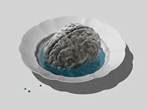
Background imageNeuroscience Collection: Donor brain, conceptual artwork

Donor brain, conceptual artwork
Donor brain on a dish, conceptual computer artwork

Background imageNeuroscience Collection: Circulatory system and brain, artwork

Circulatory system and brain, artwork
Circulatory system and brain. Computer artwork showing the blood vessels of the head and neck. Also seen is the brain (yellow)

Background imageNeuroscience Collection: Cardiovascular system and brain, artwork

Cardiovascular system and brain, artwork
Cardiovascular system and brain. Computer artwork showing the blood vessels of the torso, arms, head and neck. Also seen is the brain (yellow)

Background imageNeuroscience Collection: Neural network, abstract artwork

Neural network, abstract artwork
Neural network. Abstract computer artwork representing nerve cells, or neurons. Neurons are responsible for passing information around the central nervous system (CNS)

Background imageNeuroscience Collection: Neural network, computer artwork

Neural network, computer artwork
Neural network. Conceptual computer artwork of a brain, with the brains neural network represented by blue strands in the background. The front of the brain is at right

Background imageNeuroscience Collection: Myelinated nerves, SEM

Myelinated nerves, SEM
Myelinated nerves. Coloured scanning electron micrograph (SEM) of a section through myelinated nerve fibres and Schwann cells

Background imageNeuroscience Collection: Glial cells, SEM

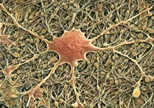
Glial cells, SEM
Glial cells. Coloured scanning electron micrograph (SEM) of a glial cell (centre). Glial cells are nervous system cells that provide structural support and protection for neurons (nerve cells)

Background imageNeuroscience Collection: Norepinephrine neurotransmitter molecule

Norepinephrine neurotransmitter molecule. Computer model showing the structure of the neurotransmitter and hormone norepinephrine, or noradrenaline

Background imageNeuroscience Collection: Met-enkephalin molecule

Met-enkephalin molecule. Computer model showing the structure of the neurotransmitter met-enkephalin (methionine-enkephalin)


All Professionally Made to Order for Quick Shipping

"Unraveling the Mysteries of the Mind: Exploring Neuroscience through Art and Science" Delve into the intricate world of neuroscience, where art meets science to unravel the complexities of the human brain. Starting with an inferior view, witness a captivating portrayal of its anatomy, showcasing the mesmerizing Medulla Oblongata – a vital structure responsible for controlling essential bodily functions. Moving to a superior view, be enchanted by colored lobes and labels that demystify different regions within our brain. Marvel at how these distinct areas work in harmony to shape our thoughts, emotions, and actions. Prepare to question your perception as you encounter mind-bending artwork like the Hollow-face illusion. Witness how our brains can be deceived by optical tricks, highlighting their remarkable ability to interpret reality. Venturing further into lateral views of human brain anatomy reveals another fascinating aspect – glial cells. Observe them through confocal light micrography; these unsung heroes play crucial roles in supporting neurons and maintaining overall brain health. Returning once more to artistic interpretations, explore additional illusions such as Ouchi illusion and Goblet illusion. These captivating artworks challenge our understanding of visual perception while reminding us that our brains are constantly processing information in unique ways. But it's not all about illusions; we must also acknowledge the importance of blood supply within brain tissue. Discover how this intricate network ensures oxygenation and nourishment for optimal cognitive function. Concluding this journey is yet another stunning artwork depicting various aspects of brain anatomy. Reflect on what you have learned throughout this exploration - from structural intricacies to perceptual wonders - gaining a deeper appreciation for neuroscience's ongoing quest to understand one of humanity's greatest enigmas.