Fraud Blocker Skip to main content

Neuroscience Collection (page 3)

"Unraveling the Mysteries of the Mind: Exploring Neuroscience through Art and Science" Delve into the intricate world of neuroscience

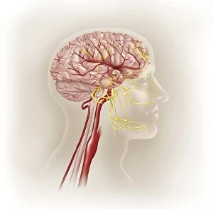
Background imageNeuroscience Collection: Detail of ateries of the human head and the trigeminal nerve

Detail of ateries of the human head and the trigeminal nerve

Background imageNeuroscience Collection: Microscopic view of inner nervous system

Microscopic view of inner nervous system

Background imageNeuroscience Collection: Microscopic view of a multipolar neuron

Microscopic view of a multipolar neuron. Multipolar neurons possess a single axon and many dendrites

Background imageNeuroscience Collection: Illustration of action potential of a nerve cell

Illustration of action potential of a nerve cell

Background imageNeuroscience Collection: Conceptual image of a neuron

Conceptual image of a neuron

Background imageNeuroscience Collection: Directed differentiation of multipotential human neural progenitor cells

Directed differentiation of multipotential human neural progenitor cells
Human neural progenitor cells were isolated under selective culture conditions from the developing human brain and directed through lineage differentiation to GFAP + (glial fibrillary acid protein)

Background imageNeuroscience Collection: Human brain anatomy, lateral view, with labels

Human brain anatomy, lateral view, with labels

Background imageNeuroscience Collection: Anatomy of human skull, eyeball and arteries to brain

Anatomy of human skull, eyeball and arteries to brain

Background imageNeuroscience Collection: Prion disease treatment, artwork C018 / 1838

Prion disease treatment, artwork C018 / 1838
Prion disease treatment. Artwork of three pyramidal neurons (nerve cells). The cell at left is healthy. The cell at back has died because of a prion (pink) infection

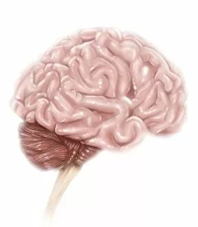
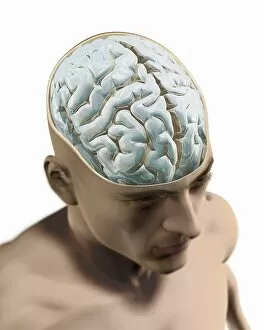
Background imageNeuroscience Collection: Human brain, artwork F006 / 6313

Human brain, artwork F006 / 6313
Human brain, computer artwork. The front of the brain is at bottom

Background imageNeuroscience Collection: Neural progenitor cell differentiation C018 / 8758

Neural progenitor cell differentiation C018 / 8758
Neural progenitor cell differentiation. Fluorescence light micrograph of neural progenitor cells that have been grown in a medium that is selective for astrocytes for three weeks

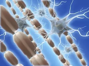
Background imageNeuroscience Collection: Myelin sheats and glial cells, artwork C014 / 2646

Myelin sheats and glial cells, artwork C014 / 2646
Computer artwork depicting axons surrounded by a myelin sheath (brown) and microglial cells (light blue). Glial cells are nervous system cells that provide structural support

Background imageNeuroscience Collection: Stem cell-derived neurons, micrograph

Stem cell-derived neurons, micrograph
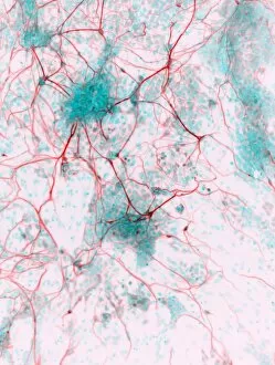
Stem cell-derived neurons. Light micrograph of human nerve cells (neurons) that have been derived from induced pluripotent stem cells (IPS). Tuj1 proteins are cyan, and cell nuclei are red

Background imageNeuroscience Collection: Stem cell-derived astrocyte brain cells

Stem cell-derived astrocyte brain cells
Stem cell-derived nerve cells. Fluorescence light micrograph of astrocyte brain cells that have been derived from neural (nerve) stem cells from a mouse

Background imageNeuroscience Collection: Effect of Parkinsons disease, artwork

Effect of Parkinsons disease, artwork
Effect of Parkinsons disease. Computer artwork showing the neural pathways within the brain that are affected by a lack of dopamine, caused by the onset of Parkinsons disease

Background imageNeuroscience Collection: Peripheral nerve, light micrograph

Peripheral nerve, light micrograph
Peripheral nerve. Light micrograph of a section through a peripheral nerve. This is a mixed nerve with myelinated axons (dark blue circles)

Background imageNeuroscience Collection: Nerve ganglion, light micrograph

Nerve ganglion, light micrograph
Nerve ganglion. Light micrograph of a section through a dorsal (sensory) spinal root ganglion associated with a sensory nerve root of the spinal cord. Sensory information from peripheral sites e.g

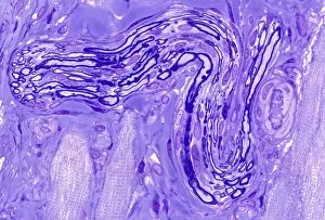
Background imageNeuroscience Collection: Myelinated nerve, light micrograph

Myelinated nerve, light micrograph
Myelinated nerve. Light micrograph of a section through a peripheral myelinated nerve, showing many individual axons each covered with deep blue-staining myelin sheaths

Background imageNeuroscience Collection: Acetylcholine receptor-conotoxin complex

Acetylcholine receptor-conotoxin complex. Molecular model showing alpha-conotoxin bound to a nicotinic acetlycholine receptor

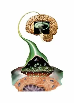
Background imageNeuroscience Collection: Brain synapse, anatomical artwork

Brain synapse, anatomical artwork
Brain synapse. Anatomical computer artwork of a human brain with an enlargement showing the structure of a synapse (lower right) within one of the striate bodies that make up the striatum

Background imageNeuroscience Collection: Striatum, anatomical artwork

Striatum, anatomical artwork
Striatum. Anatomical computer artwork of a human brain, showing the striate bodies (blue and green) in the midbrain, and the substantia nigra (black) in the brainstem

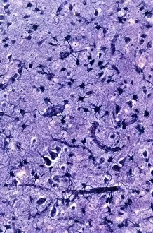
Background imageNeuroscience Collection: Glial cells, light micrograph

Glial cells, light micrograph
Glial cells in the brain. Light micrograph of a section through glial cells (dark) in the grey matter of the brain. Due to their star shape these glial cells are called astrocytes

Background imageNeuroscience Collection: Nerve cell degeneration, artwork

Nerve cell degeneration, artwork
Nerve cell degeneration. Computer artwork showing a collection of nerve cells (neurons) that have become damaged

Background imageNeuroscience Collection: Limbic system of the brain, artwork C015 / 4529

Limbic system of the brain, artwork C015 / 4529
Limbic system of the brain. Computer artwork showing the main parts of the limbic system from the human brain. At centre is the oval-shaped thalamus (red) which receives sensory stimuli

Background imageNeuroscience Collection: Hearing centre, artwork

Hearing centre, artwork
Hearing centre. Computer artwork showing the area of the brains temporal lobe that is associated with hearing (red). This area receives auditory input from the cochlea (coiled)

Background imageNeuroscience Collection: Human brain, anatomical artwork

Human brain, anatomical artwork
Human brain. Anatomical computer artwork of a human brain seen from below, showing the cranial nerves (centre)

Background imageNeuroscience Collection: Substantia nigra, artwork

Substantia nigra, artwork
Substantia nigra. Computer artwork showing a healthy substantia nigra (left) and degenerated substantia nigra (right) from human brains

Background imageNeuroscience Collection: Photic sneeze reflex, artwork C016 / 7700

Photic sneeze reflex, artwork C016 / 7700
Photic sneeze reflex. Computer artwork representing one explanation for the photic sneeze reflex. About a quarter of the population sneeze when they look at a bright light

Background imageNeuroscience Collection: Synapse, artwork C016 / 7535

Synapse, artwork C016 / 7535
Synapse. Computer artwork of a synapse, the junction between nerve cells (orange). Synapses transmit electrical signals from one nerve cell to the next

Background imageNeuroscience Collection: Human brain, artwork C016 / 9370

Human brain, artwork C016 / 9370
Human brain, computer artwork

Background imageNeuroscience Collection: Brain glial cells, light micrograph C016 / 0524

Brain glial cells, light micrograph C016 / 0524
Brain glial cells. Light micrograph of a section through brain tissue, showing numerous glial cells (dark). Glial cells, or neuroglia, are non-neural cells of the central nervous system (CNS)

Background imageNeuroscience Collection: Brain glial cells, light micrograph C016 / 0523

Brain glial cells, light micrograph C016 / 0523
Brain glial cells. Light micrograph of a section through brain tissue, showing numerous glial cells (dark). Glial cells, or neuroglia, are non-neural cells of the central nervous system (CNS)

Background imageNeuroscience Collection: C. elegans worm, illustration C018 / 0731

C. elegans worm, illustration C018 / 0731
Caenorhabditis elegans worm, illustration. C. elegans is a soil-dwelling hermaphrodite nematode worm and one of the most studied animals in biological and genetic research

Background imageNeuroscience Collection: Brain areas, conceptual illustration C018 / 0749

Brain areas, conceptual illustration C018 / 0749
Brian areas, conceptual illustration. Different areas of the cerebral cortex are involved in different cognitive and behavioural tasks

Background imageNeuroscience Collection: Nervous system cells, artwork C017 / 3423

Nervous system cells, artwork C017 / 3423
Nervous system cells. Computer artwork showing neurons (nerve cells, purple), astrocytes (green) and a blood vessel (along right)

Background imageNeuroscience Collection: Myelin sheats and glial cells, artwork C014 / 2647

Myelin sheats and glial cells, artwork C014 / 2647
Computer artwork depicting axons surrounded by a myelin sheath (brown) and microglial cells (light blue). Glial cells are nervous system cells that provide structural support

Background imageNeuroscience Collection: Induced nerve stem cells, micrograph

Induced nerve stem cells, micrograph
Induced nerve stem cells. Fluorescence light micrograph of neural (nerve) stem cells that have been created (induced) from human adult skin fibroblast cells by gene manipulation

Background imageNeuroscience Collection: Stem cell-derived nerve cells

Stem cell-derived nerve cells. Fluorescence light micrograph of neural (nerve) stem cells that have been derived from human embryonic stem cells (HESC)

Background imageNeuroscience Collection: Brain pacemaker, artwork

Brain pacemaker, artwork
Brain pacemaker. Computer artwork showing a deep brain stimulation (DBS) electrode (right and inset) implanted in a persons brain, with a control box located on the persons chest (lower left)

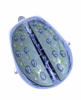
Background imageNeuroscience Collection: EEG, electrodes on head, artwork

EEG, electrodes on head, artwork
Electroencephalography. Computer artwork depicting of a EEG cap on top of a head, showing the electrodes attached to the scalp. EEG measures and records the electrical activity of the brain

Background imageNeuroscience Collection: Neuron firing, conceptual artwork C013 / 5649

Neuron firing, conceptual artwork C013 / 5649
Neuron firing, conceptual computer artwork

Background imageNeuroscience Collection: Visual processes, conceptual artwork

Visual processes, conceptual artwork


All Professionally Made to Order for Quick Shipping

"Unraveling the Mysteries of the Mind: Exploring Neuroscience through Art and Science" Delve into the intricate world of neuroscience, where art meets science to unravel the complexities of the human brain. Starting with an inferior view, witness a captivating portrayal of its anatomy, showcasing the mesmerizing Medulla Oblongata – a vital structure responsible for controlling essential bodily functions. Moving to a superior view, be enchanted by colored lobes and labels that demystify different regions within our brain. Marvel at how these distinct areas work in harmony to shape our thoughts, emotions, and actions. Prepare to question your perception as you encounter mind-bending artwork like the Hollow-face illusion. Witness how our brains can be deceived by optical tricks, highlighting their remarkable ability to interpret reality. Venturing further into lateral views of human brain anatomy reveals another fascinating aspect – glial cells. Observe them through confocal light micrography; these unsung heroes play crucial roles in supporting neurons and maintaining overall brain health. Returning once more to artistic interpretations, explore additional illusions such as Ouchi illusion and Goblet illusion. These captivating artworks challenge our understanding of visual perception while reminding us that our brains are constantly processing information in unique ways. But it's not all about illusions; we must also acknowledge the importance of blood supply within brain tissue. Discover how this intricate network ensures oxygenation and nourishment for optimal cognitive function. Concluding this journey is yet another stunning artwork depicting various aspects of brain anatomy. Reflect on what you have learned throughout this exploration - from structural intricacies to perceptual wonders - gaining a deeper appreciation for neuroscience's ongoing quest to understand one of humanity's greatest enigmas.