Fraud Blocker Skip to main content

Neuroscience Collection (page 5)

"Unraveling the Mysteries of the Mind: Exploring Neuroscience through Art and Science" Delve into the intricate world of neuroscience

Background imageNeuroscience Collection: Neuropeptide Y neurotransmitter molecule C015 / 6240

Neuropeptide Y neurotransmitter molecule C015 / 6240
Neuropeptide Y neurotransmitter molecule. Molecular model showing the structure of the neurotransmitter neuropeptide Y (NPY). NPY is found in the brain and autonomic nervous system

Background imageNeuroscience Collection: Myelinated nerve, TEM C016 / 5840

Myelinated nerve, TEM C016 / 5840
Myelinated nerve. Coloured transmission electron micrograph (TEM) of a section through a myelinated nerve fibre and Schwann cell

Background imageNeuroscience Collection: Myelinated nerve, TEM C016 / 5839

Myelinated nerve, TEM C016 / 5839
Myelinated nerve. Coloured transmission electron micrograph (TEM) of a section through a myelinated nerve fibre and Schwann cell

Background imageNeuroscience Collection: Myelinated nerve, TEM C016 / 5838

Myelinated nerve, TEM C016 / 5838
Myelinated nerve. Coloured transmission electron micrograph (TEM) of a section through a myelinated nerve fibre and Schwann cell

Background imageNeuroscience Collection: Myelinated nerve, TEM C016 / 5448

Myelinated nerve, TEM C016 / 5448
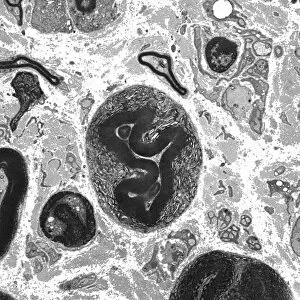
Myelinated nerve. Transmission electron micrograph (TEM) of a section through a myelinated nerve fibre and Schwann cell. Myelin (black)

Background imageNeuroscience Collection: Myelinated nerve, TEM C016 / 5370

Myelinated nerve, TEM C016 / 5370
Myelinated nerve. Transmission electron micrograph (TEM) of a section through a myelinated nerve fibre and Schwann cell (centre)

Background imageNeuroscience Collection: Neuropeptide Y neurotransmitter molecule C014 / 0013

Neuropeptide Y neurotransmitter molecule C014 / 0013
Neuropeptide Y neurotransmitter molecule. Molecular model showing the structure of the neurotransmitter neuropeptide Y (NPY)

Background imageNeuroscience Collection: Synapses, artwork C014 / 0002

Synapses, artwork C014 / 0002
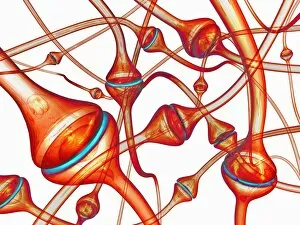
Synapses. Computer artwork of synapses, the junction between nerve cells (orange). Synapses transmit electrical signals from one nerve cell to the next
Background imageNeuroscience Collection: Synapses, artwork C014 / 0004

Synapses, artwork C014 / 0004
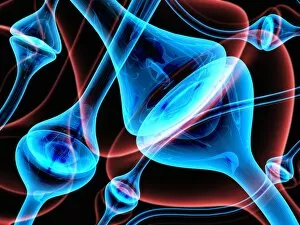
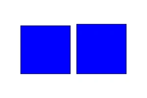
Synapses. Computer artwork of synapses, the junction between nerve cells (blue). Synapses transmit electrical signals from one nerve cell to the next

Background imageNeuroscience Collection: Synapses, artwork C014 / 0003

Synapses, artwork C014 / 0003
Synapses. Computer artwork of synapses, the junction between nerve cells (blue). Synapses transmit electrical signals from one nerve cell to the next

Background imageNeuroscience Collection: Myelinated nerves, SEM C013 / 7142

Myelinated nerves, SEM C013 / 7142
Myelinated nerves. Coloured scanning electron micrograph (SEM) of a section through the sciatic nerve, showing the myelinated nerve fibres (axons)

Background imageNeuroscience Collection: Myelinated nerves, SEM C013 / 7141

Myelinated nerves, SEM C013 / 7141
Myelinated nerves. Coloured scanning electron micrograph (SEM) of a section through the sciatic nerve, showing the myelinated nerve fibres (axons)

Background imageNeuroscience Collection: Myelinated nerves, SEM C013 / 7138

Myelinated nerves, SEM C013 / 7138
Myelinated nerves. Coloured scanning electron micrograph (SEM) of a section through a myelinated nerve fibre (axon, beige, centre) from the sciatic nerve

Background imageNeuroscience Collection: Ciliary hairs in the cerebral ventricle

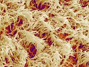
Ciliary hairs in the cerebral ventricle. Coloured scanning electron micrograph (SEM) of the wall of a cerebral ventricle. The cerebral ventricles are four communicating cavities within the brain that

Background imageNeuroscience Collection: Brain anatomy, 14th century artwork

Brain anatomy, 14th century artwork. The cranial vault (top of the skull, left) has been removed and two of the skull sutures (zig-zag lines) are shown

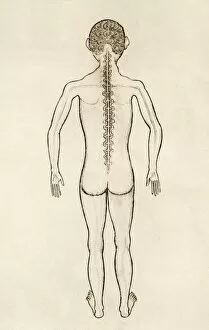
Background imageNeuroscience Collection: Spinal anatomy, 14th century artwork

Spinal anatomy, 14th century artwork. The spinal cord is shown running down the back, with eighteen spinal nerves braching off through holes in the vertebrae (spinal bones)

Background imageNeuroscience Collection: Impossible shape

Impossible shape, computer artwork

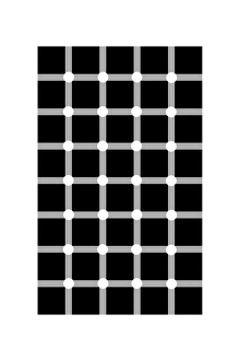
Background imageNeuroscience Collection: Scintillating grid illusion

Scintillating grid illusion. This illusion, based on the Hermann grid, was created in 1994. When the grid is observed, the white dots at the intersection of the grey lines appear to scintillate

Background imageNeuroscience Collection: Square illusion - subdivision

Square illusion - subdivision leads to apparent enlargement. There are three squares of identical area. However, the middle square appears to be wider than it is tall

Background imageNeuroscience Collection: Muller-Lyer illusion

Muller-Lyer illusion. The two lines with arrows at each end appear to be different lengths on the left. In fact they are the same length

Background imageNeuroscience Collection: Impossible rectangle

Impossible rectangle. This is an impossible object - one that can be drawn in 2 dimensions but not created

Background imageNeuroscience Collection: Illusory contours

Illusory contours forming circles. This illusion demonstrates the false perception of areas of increased brightness. Lines are apparently joined to bright circles. In fact no such circles exist

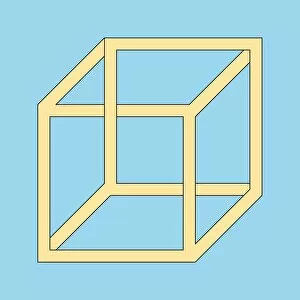
Background imageNeuroscience Collection: Freemish crate

Freemish crate. This representation of an impossible cube is based upon the standard 2- dimensional depiction of a cube, but with some of the edges crossing in a way that could not be achieved in 3

Background imageNeuroscience Collection: Jastrow illusion

Jastrow illusion. The bottom shape appears to be larger than the top shape. They are actually identical

Background imageNeuroscience Collection: Circle in a square illusion

Circle in a square illusion. The two red circles in the image are the same size. However, the one inside a blue square looks larger than the one enclosing a blue square

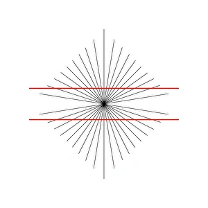
Background imageNeuroscience Collection: Hering illusion

Hering illusion. This illusion was created by the physiologist Edward Hering in the 19th century. It exploits the brains understanding of perspective to fool it into thinking that the two horizontal

Background imageNeuroscience Collection: Bullseye illusion

Bullseye illusion. The blue shaded area in the centre of the circle appears larger than the blue perimeter area. In fact they are the same area. This is easily proven by calculation

Background imageNeuroscience Collection: Framing colour illusion

Framing colour illusion
Framing colours illusion. Black borders around colours enhance their brightness and prevent clashing. The colours in the pattern on the left appear to be brighter and bolder than those on the right

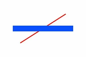
Background imageNeuroscience Collection: Poggendorff illusion

Poggendorff illusion. The two red lines crossing the blue bar are colinear. Many people see them as being offset. There are still no satisfactory explanations as to why this should be the case

Background imageNeuroscience Collection: Three arc illusion

Three arc illusion. The three arcs shown give the impression of belonging to circles of increasing radius when viewed from top to bottom. They actually come from circles of the same radius

Background imageNeuroscience Collection: Square illusion - orientation

Square illusion - orientation changes perception of size. The two squares are identical. However, the one on the left appears larger because it has been rotated through 45 degrees

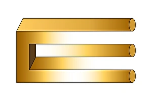
Background imageNeuroscience Collection: Impossible trident

Impossible trident. This uses the fact that a rectangular bar can be represented in 2 dimensions by 3 lines, whereas a cylinder requires 2. So we have 3 cylinders blending into 2 rectangular bars

Background imageNeuroscience Collection: Kanizsa triangle

Kanizsa triangle. This image appears to depict a solid triangle overlaying another triangle. This triangle looks brighter than the background. In fact there is no triangle

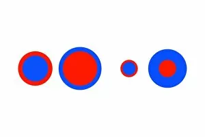
Background imageNeuroscience Collection: Delboeuf illusion

Delboeuf illusion. An illusion created by the Belgian philosopher Franz Joseph Delboeuf. The two red circles on the left have the same diameter, as do the two red circles on the right

Background imageNeuroscience Collection: Computer artwork of an impossible triangle

Computer artwork of an impossible triangle
Impossible triangle. Abstract computer artwork of a triangle, made from 3 sections of tower block, which would be impossible to construct because it breaks the laws of artistic perspective

Background imageNeuroscience Collection: Angle illusion

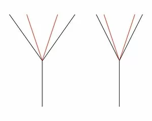
Angle illusion. The red angle marked on the left appears to be larger than the red angle on the right. In fact they are the same size

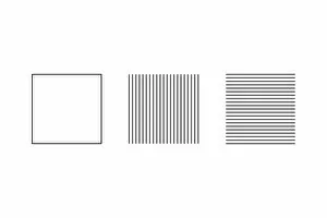
Background imageNeuroscience Collection: Square illusion - vertical lines appear longer

Square illusion - vertical lines appear longer
Square illusion. The shape on the right hand side is a square whilst the one on the left, which is slightly shorter, is not. However many people believe it to be the other way round

Background imageNeuroscience Collection: Orbison illusion

Orbison illusion. Both the rectangle surrounding the pattern and the perfect square within the pattern appear to be distorted

Background imageNeuroscience Collection: Irradiation illusion

Irradiation illusion. The white square against a black background on the left appears to be larger than the black square against a white background on the right. They are actually the same size

Background imageNeuroscience Collection: Cafe wall illusion

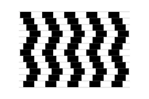
Cafe wall illusion. This illusion is so named because it was first noticed on a tiled wall in a Bristol cafe. The pattern in the image appears to show sloping grey lines between the black

Background imageNeuroscience Collection: Simultaneous contrast

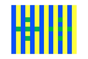
Simultaneous contrast example. The green squares look brighter on the left, where they are in yellow bands, compared to the right, where they are in blue bands

Background imageNeuroscience Collection: Zoellner illusion

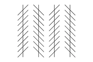
Zoellner illusion. The vertical lines are parallel but seem to be tilted. There are 2 theories as to why this is the case

Background imageNeuroscience Collection: Hermann-Hering illusion

Hermann-Hering illusion
Hermann grid illusion. The intersections of the white horizontal and vertical lines in this image appear to be grey. But when looked at directly they are clearly white

Background imageNeuroscience Collection: Penrose stairway

Penrose stairway. This is an impossible figure created by the physicist Roger Penrose and used by M C Escher in his illustration " Ascending and Descending"

Background imageNeuroscience Collection: Ehrenstein illusion

Ehrenstein illusion - illusory contours forming circles. Created in 1954 by the psychologist Walter Ehrenstein, this illusion demonstrates the false perception of areas of increased brightness

Background imageNeuroscience Collection: Brain-computer interface, artwork

Brain-computer interface, artwork
Brain-computer interface. Artwork showing a paralysed man using his thoughts to play a computer game. This setup is similar to that used by Matthew Nagle

Background imageNeuroscience Collection: Brain function, conceptual artwork

Brain function, conceptual artwork. Electroencephalogram (EEG) machine monitoring brain activity

Background imageNeuroscience Collection: Nerve cells, light micrograph

Nerve cells, light micrograph
Nerve cells, or neurons, light micrograph. Neurons are responsible for passing information around the central nervous system (CNS) and from the CNS to the rest of the body


All Professionally Made to Order for Quick Shipping

"Unraveling the Mysteries of the Mind: Exploring Neuroscience through Art and Science" Delve into the intricate world of neuroscience, where art meets science to unravel the complexities of the human brain. Starting with an inferior view, witness a captivating portrayal of its anatomy, showcasing the mesmerizing Medulla Oblongata – a vital structure responsible for controlling essential bodily functions. Moving to a superior view, be enchanted by colored lobes and labels that demystify different regions within our brain. Marvel at how these distinct areas work in harmony to shape our thoughts, emotions, and actions. Prepare to question your perception as you encounter mind-bending artwork like the Hollow-face illusion. Witness how our brains can be deceived by optical tricks, highlighting their remarkable ability to interpret reality. Venturing further into lateral views of human brain anatomy reveals another fascinating aspect – glial cells. Observe them through confocal light micrography; these unsung heroes play crucial roles in supporting neurons and maintaining overall brain health. Returning once more to artistic interpretations, explore additional illusions such as Ouchi illusion and Goblet illusion. These captivating artworks challenge our understanding of visual perception while reminding us that our brains are constantly processing information in unique ways. But it's not all about illusions; we must also acknowledge the importance of blood supply within brain tissue. Discover how this intricate network ensures oxygenation and nourishment for optimal cognitive function. Concluding this journey is yet another stunning artwork depicting various aspects of brain anatomy. Reflect on what you have learned throughout this exploration - from structural intricacies to perceptual wonders - gaining a deeper appreciation for neuroscience's ongoing quest to understand one of humanity's greatest enigmas.