Fraud Blocker Skip to main content

Neurology Collection (page 5)

Neurology, the fascinating study of the nervous system, unravels the intricate workings of our brain and its various components

Background imageNeurology Collection: Figure 10: Showing the expressive lines of m. frontalis in a young girl, 1854-56

Figure 10: Showing the expressive lines of m. frontalis in a young girl, 1854-56, printed 1862

Background imageNeurology Collection: Figure 55: Astonishment badly rendered by the subject: a ridiculous and i

Figure 55: Astonishment badly rendered by the subject: a ridiculous and inane expression. 1854-56, printed 1862

Background imageNeurology Collection: Figure 21: Painful recollection and recollection or calling something to

Figure 21: Painful recollection and recollection or calling something to mind, 1854-56, printed 1862

Background imageNeurology Collection: Figure 71: Same head as in Plate 70, 1854-56, printed 1862. Creators: Duchenne de Boulogne

Figure 71: Same head as in Plate 70, 1854-56, printed 1862. Creators: Duchenne de Boulogne, Adrien Alban Tournachon
Figure 71: Same head as in Plate 70, 1854-56, printed 1862

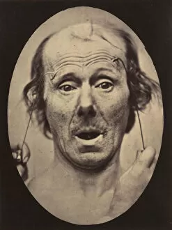
Background imageNeurology Collection: Figure 57: Astonishment, stupefaction, amazement, 1854-56, printed 1862

Figure 57: Astonishment, stupefaction, amazement, 1854-56, printed 1862

Background imageNeurology Collection: Figure 73: Head of Niobe, 1854-56, printed 1862. Creators: Duchenne de Boulogne

Figure 73: Head of Niobe, 1854-56, printed 1862. Creators: Duchenne de Boulogne
Figure 73: Head of Niobe, 1854-56, printed 1862

Background imageNeurology Collection: Figure 12: A study of the contraction of and the expression produced by t

Figure 12: A study of the contraction of and the expression produced by the superior part of m. orbicularis oculi, 1854-56, printed 1862

Background imageNeurology Collection: Figure 47: A suggestion of this same weeping, 1854-56, printed 1862

Figure 47: A suggestion of this same weeping, 1854-56, printed 1862

Background imageNeurology Collection: Figure 56: Surprise, 1854-56, printed 1862. Creators: Duchenne de Boulogne

Figure 56: Surprise, 1854-56, printed 1862. Creators: Duchenne de Boulogne
Figure 56: Surprise, 1854-56, printed 1862

Background imageNeurology Collection: Figure 63: Expression of terror, 1854-56, printed 1862. Creators: Duchenne de Boulogne

Figure 63: Expression of terror, 1854-56, printed 1862. Creators: Duchenne de Boulogne
Figure 63: Expression of terror, 1854-56, printed 1862

Background imageNeurology Collection: Figure 34: Grimace, 1854-56, printed 1862. Creators: Duchenne de Boulogne

Figure 34: Grimace, 1854-56, printed 1862. Creators: Duchenne de Boulogne
Figure 34: Grimace, 1854-56, printed 1862

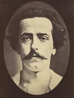
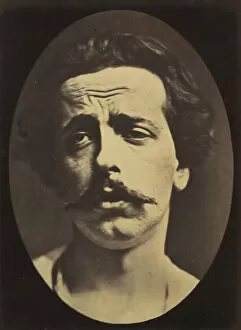
Background imageNeurology Collection: Figure 4: The face in repose of a young man, 1854-56, printed 1862

Figure 4: The face in repose of a young man, 1854-56, printed 1862

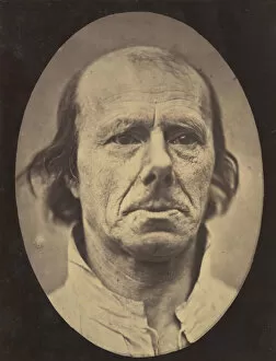
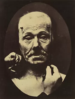
Background imageNeurology Collection: Figure 3: The face of an old man... photographed in repose. 1854-56, printed 1862

Figure 3: The face of an old man... photographed in repose. 1854-56, printed 1862

Background imageNeurology Collection: Figure 83: Lady Macbeth, ferocious cruelty, 1854-56, printed 1862

Figure 83: Lady Macbeth, ferocious cruelty, 1854-56, printed 1862

Background imageNeurology Collection: Figure 82: Lady Macbeth, strong expression of cruelty, 1854-56, printed 1862

Figure 82: Lady Macbeth, strong expression of cruelty, 1854-56, printed 1862

Background imageNeurology Collection: Figure 8: Contraction of the right m. frontalis. 1854-56, printed 1862

Figure 8: Contraction of the right m. frontalis. 1854-56, printed 1862

Background imageNeurology Collection: Figure 24: Extreme pain to the point of exhaustion, the head of Christ an

Figure 24: Extreme pain to the point of exhaustion, the head of Christ and memory of love or ecstatic gaze. 1854-56, printed 1862

Background imageNeurology Collection: Icono-photographique. Mecanisme de la Physionomie Humaine. Fig

Icono-photographique. Mecanisme de la Physionomie Humaine. Fig. 65, 1854-56, printed 1862

Background imageNeurology Collection: Figure 19: Suffering, 1854-56, printed 1862. Creators: Duchenne de Boulogne

Figure 19: Suffering, 1854-56, printed 1862. Creators: Duchenne de Boulogne
Figure 19: Suffering, 1854-56, printed 1862

Background imageNeurology Collection: Figure 48: Mild weeping, pity and feeble false laughter, 1854-56, printed 1862

Figure 48: Mild weeping, pity and feeble false laughter, 1854-56, printed 1862

Background imageNeurology Collection: Figure 25: Not an expression of pain, 1854-56, printed 1862

Figure 25: Not an expression of pain, 1854-56, printed 1862

Background imageNeurology Collection: Faradisation du muscle frontal, 1854-56, printed 1862. Creators: Duchenne de Boulogne

Faradisation du muscle frontal, 1854-56, printed 1862. Creators: Duchenne de Boulogne
Faradisation du muscle frontal, 1854-56, printed 1862

Background imageNeurology Collection: Figure 52: Voluntary retraction of the lower lip, 1854-56, printed 1862

Figure 52: Voluntary retraction of the lower lip, 1854-56, printed 1862

Background imageNeurology Collection: Figure 54: Voluntary lowering of the lower jaw, 1854-56, printed 1862

Figure 54: Voluntary lowering of the lower jaw, 1854-56, printed 1862

Background imageNeurology Collection: Figure 66: Head of Arrotino (the spy, the knife grinder, and so on), 1854-56

Figure 66: Head of Arrotino (the spy, the knife grinder, and so on), 1854-56, printed 1862

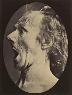
Background imageNeurology Collection: Figure 22: No painful expression, 1854-56, printed 1862

Figure 22: No painful expression, 1854-56, printed 1862

Background imageNeurology Collection: Figure 49: Painful weeping and forward looking. 1854-56, printed 1862

Figure 49: Painful weeping and forward looking. 1854-56, printed 1862

Background imageNeurology Collection: Figure 18: Aggression, wickedness, 1854-56, printed 1862

Figure 18: Aggression, wickedness, 1854-56, printed 1862

Background imageNeurology Collection: Figure 17: On the right, electrization of m. procerus: severity, aggressi

Figure 17: On the right, electrization of m. procerus: severity, aggression. On the left: attention. 1854-56, printed 1862

Background imageNeurology Collection: Figure 60: Fright, 1854-56, printed 1862. Creators: Duchenne de Boulogne

Figure 60: Fright, 1854-56, printed 1862. Creators: Duchenne de Boulogne
Figure 60: Fright, 1854-56, printed 1862

Background imageNeurology Collection: Figure 70: Head of the Laocoon of Rome, 1854-56, printed 1862

Figure 70: Head of the Laocoon of Rome, 1854-56, printed 1862. Creators: Duchenne de Boulogne, Adrien Alban Tournachon
Figure 70: Head of the Laocoon of Rome, 1854-56, printed 1862

Background imageNeurology Collection: Figure 45: Pain and despair. 1854-56, printed 1862. Creators: Duchenne de Boulogne

Figure 45: Pain and despair. 1854-56, printed 1862. Creators: Duchenne de Boulogne
Figure 45: Pain and despair. 1854-56, printed 1862

Background imageNeurology Collection: Figure 6: The grimace produced is similar to a tic of the face, 1854-56, printed 1862

Figure 6: The grimace produced is similar to a tic of the face, 1854-56, printed 1862

Background imageNeurology Collection: Figure 75: Nun saying her prayers, 1854-56, printed 1862

Figure 75: Nun saying her prayers, 1854-56, printed 1862

Background imageNeurology Collection: Figure 62: Terror, semiprofile, 1854-56, printed 1862. Creators: Duchenne de Boulogne

Figure 62: Terror, semiprofile, 1854-56, printed 1862. Creators: Duchenne de Boulogne
Figure 62: Terror, semiprofile, 1854-56, printed 1862

Background imageNeurology Collection: Figure 78: Scene of coquetry, 1854-56, printed 1862. Creators: Duchenne de Boulogne

Figure 78: Scene of coquetry, 1854-56, printed 1862. Creators: Duchenne de Boulogne
Figure 78: Scene of coquetry, 1854-56, printed 1862

Background imageNeurology Collection: Figure 16: Expression of severity, 1854-56, printed 1862

Figure 16: Expression of severity, 1854-56, printed 1862

Background imageNeurology Collection: Figure 50: Affected weeping and face in repose, 1854-56, printed 1862

Figure 50: Affected weeping and face in repose, 1854-56, printed 1862

Background imageNeurology Collection: Figure 36: Scornful laughter and scornful disgust, 1854-56, printed 1862

Figure 36: Scornful laughter and scornful disgust, 1854-56, printed 1862

Background imageNeurology Collection: Figure 20: Profound suffering, with resignation, 1854-56, printed 1862

Figure 20: Profound suffering, with resignation, 1854-56, printed 1862

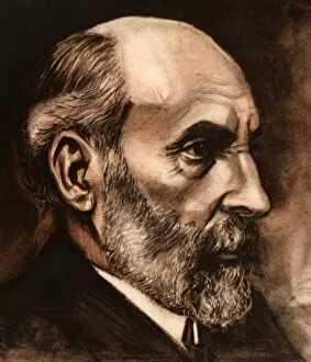
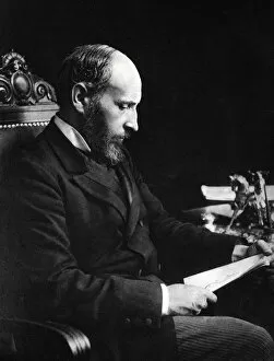
Background imageNeurology Collection: Ramon y Cajal, Santiago (Petilla of Aragon, Navarra, 1852-Madrid, 1934)

Ramon y Cajal, Santiago (Petilla of Aragon, Navarra, 1852-Madrid, 1934). Professor of Medicine and Surgery. Spanish histologist and pathologist

Background imageNeurology Collection: Santiago Ramon y Cajal (Petilla of Aragon, Navarra, 1852-Madrid, 1934), Professor

Santiago Ramon y Cajal (Petilla of Aragon, Navarra, 1852-Madrid, 1934), Professor of Medicine and Surgery, histologist and pathologist

Background imageNeurology Collection: Albrecht von Haller, 18th century Swiss physician and scientist, c1840

Albrecht von Haller, 18th century Swiss physician and scientist, c1840
Albrecht von Haller, 18th century Swiss physician and scientist, [c1840]. Von Haller (1708-1777) worked in the field of experimental physiology and was the founder of the science of neurology

Background imageNeurology Collection: Albrecht von Haller, Swiss physician and scientist, c1770 (c1780)

Albrecht von Haller, Swiss physician and scientist, c1770 (c1780). Von Haller (1708-1777) worked in the field of experimental physiology and was the founder of the science of neurology

Background imageNeurology Collection: Jean Martin Charcot demonstrating hypnosis, 1879

Jean Martin Charcot demonstrating hypnosis, 1879. Charcot (1825-1893), French neurologist and pathologist, demonstrating the production of hypnosis using beam of light from a magic lantern

Background imageNeurology Collection: Jean Martin Charcot, French neurologist and pathologist, 1893

Jean Martin Charcot, French neurologist and pathologist, 1893. Charcot (1825-1893) worked at the Salpetriere Hospital, Paris from 1862, and established a neurological unit there

Background imageNeurology Collection: Charles Bell (1774-1842), Scottish surgeon and anatomist

Charles Bell (1774-1842), Scottish surgeon and anatomist. Bell made several discoveries in the field of neurology. Bells Palsy (a facial paralysis) is named after him

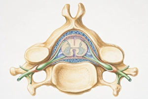
Background imageNeurology Collection: Anterior cross section of spinal column revealing how vertebra fits round spinal cord

Anterior cross section of spinal column revealing how vertebra fits round spinal cord


All Professionally Made to Order for Quick Shipping

Neurology, the fascinating study of the nervous system, unravels the intricate workings of our brain and its various components. From the Motor homunculus model that depicts how different body parts are represented in our motor cortex to the Histological Diagram of a Mammalian Retina revealing the complexity of our visual processing, every aspect is awe-inspiring. The Cerebellum tissue, as seen through a light micrograph, showcases its unique structure responsible for coordinating movement and balance. Meanwhile, an inferior view of the Anatomy of human brain highlights its remarkable organization and interconnectedness. Through Brain fibres captured by DTI MRI scans like C017 / 7099 or C017 / 7035, we gain insights into neural pathways that facilitate communication between different regions. These pathways form a complex network crucial for transmitting information throughout our brain. Exploring Brain blood vessels using a 3D angiogram (C007 / 1981) reveals their vital role in supplying oxygen and nutrients to sustain neuronal function. Additionally, understanding Motor and sensory homunculi helps us comprehend how specific areas within our brains control different bodily functions. Delving deeper into neurology brings us face-to-face with Medulla oblongata artwork depicting this critical region involved in regulating essential autonomic functions such as breathing and heart rate. Furthermore, observing Nerve and glial cells under a light microscope illustrates their diverse roles in supporting neurons' health and functionality. Finally, examining Synapse nerve junctions through TEM unveils these microscopic structures where electrical signals are transmitted from one neuron to another—a fundamental process underlying all brain activity. In essence, neurology takes us on an incredible journey deep within ourselves—unveiling mysteries at both macroscopic levels like anatomical structures or microscopically exploring cellular intricacies, and is through this exploration that we gain profound knowledge about how our brains work—the very essence of who we are as individuals.