Fraud Blocker Skip to main content

Neurology Collection (page 4)

Neurology, the fascinating study of the nervous system, unravels the intricate workings of our brain and its various components

Background imageNeurology Collection: Hysterical epilepsy, attack, epileptoid phase, illustration from

Hysterical epilepsy, attack, epileptoid phase, illustration from
REV287050 Hysterical epilepsy, attack, epileptoid phase, illustration from Iconographie Photographique de La Salpetriere by Bourneville et P

Background imageNeurology Collection: Attack of hysteria, second phase, illustration from

Attack of hysteria, second phase, illustration from
REV287040 Attack of hysteria, second phase, illustration from Iconographie Photographique de La Salpetriere by Bourneville et P

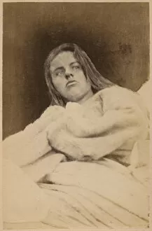
Background imageNeurology Collection: Hysterical epilepsy, after the attack, plate XXXIX from

Hysterical epilepsy, after the attack, plate XXXIX from
REV287073 Hysterical epilepsy, after the attack, plate XXXIX from Iconographie Photographique de La Salpetriere by Bourneville et P

Background imageNeurology Collection: Fig. 212 from Semiologie des Affections du Systeme Nerveux by J

Fig. 212 from Semiologie des Affections du Systeme Nerveux by J
CHT234698 Fig. 212 from Semiologie des Affections du Systeme Nerveux by J. Dejerine, 1914 (colour litho) by French School, (20th century); Bibliotheque de la Faculte de Medecine, Paris

Background imageNeurology Collection: Cross-section of the Brain, from Traite d Anatomie et de Physiologie

Cross-section of the Brain, from Traite d Anatomie et de Physiologie
CHT234693 Cross-section of the Brain, from Traite d Anatomie et de Physiologie by Felix Vicq d Azyr (1748-94) 1786 (coloured engraving) by Briceau

Background imageNeurology Collection: Jean-Martin Charcot (1825-93) August 1881 (oil on canvas)

Jean-Martin Charcot (1825-93) August 1881 (oil on canvas)
CHT234661 Jean-Martin Charcot (1825-93) August 1881 (oil on canvas) by Tofano, Eduardo (1838-1920); Musee d Histoire de la Medecine, Paris

Background imageNeurology Collection: The Structure of the Brain, from De Homine Figuris

The Structure of the Brain, from De Homine Figuris
BLT236795 The Structure of the Brain, from De Homine Figuris by Rene Descartes (1596-1650) published in The Hague, 1662 (engraving) by French School

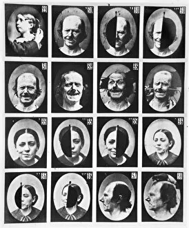
Background imageNeurology Collection: Physiognomical studies, from Mecanisme de la Physionomie humaine ou analyse

Physiognomical studies, from Mecanisme de la Physionomie humaine ou analyse
CHT207797 Physiognomical studies, from Mecanisme de la Physionomie humaine ou analyse electrophysiologique des passions by Duchenne de Boulogne, published 1876

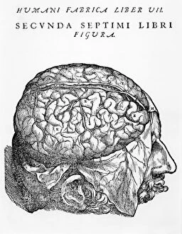
Background imageNeurology Collection: The Brain, illustration from De Humani Corporis Fabrica Librorum Epitome

The Brain, illustration from De Humani Corporis Fabrica Librorum Epitome
884588 The Brain, illustration from De Humani Corporis Fabrica Librorum Epitome by Andreas Vesalius, published in Basel, 1543 (engraving) by Vesalius

Background imageNeurology Collection: The Brain, 1825 (litho)

The Brain, 1825 (litho)
884586 The Brain, 1825 (litho) by Cloquet, Jules Germain (1790-1883); Bibliotheque de la Faculte de Medecine, Paris, France; (add.info.: Le cerveau.); eArchives Charmet; French, out of copyright

Background imageNeurology Collection: Physiognomical examination, from Mecanisme de la Physionomie humaine ou analyse

Physiognomical examination, from Mecanisme de la Physionomie humaine ou analyse
CHT207808 Physiognomical examination, from Mecanisme de la Physionomie humaine ou analyse electrophysiologique des passions by Duchenne de Boulogne, published 1876

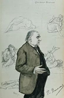
Background imageNeurology Collection: Doctor Jean Martin Charcot (1825-93) and the Hysterics, from La Revue Illustree

Doctor Jean Martin Charcot (1825-93) and the Hysterics, from La Revue Illustree
CHT227160 Doctor Jean Martin Charcot (1825-93) and the Hysterics, from La Revue Illustree, August 1887 (engraving) by Renouard, Charles Paul (1845-1924) (after); Musee de la Ville de Paris

Background imageNeurology Collection: Cross-section of the Brain, 1746 (coloured engraving)

Cross-section of the Brain, 1746 (coloured engraving)
CHT234692 Cross-section of the Brain, 1746 (coloured engraving) by Gautier d Agoty, Jacques-Fabien (1710-81); Bibliotheque de la Faculte de Medecine, Paris, France; eArchives Charmet; French

Background imageNeurology Collection: The Brain, from Traite d Anatomie de Cerveau by Felix Vicq d

The Brain, from Traite d Anatomie de Cerveau by Felix Vicq d
CHT236796 The Brain, from Traite d Anatomie de Cerveau by Felix Vicq d Azyr (1748-94) 1786 (coloured engraving) by Allais (nee Briceau)

Background imageNeurology Collection: Hysterical epilepsy, attack, crucifixion, plate IX from

Hysterical epilepsy, attack, crucifixion, plate IX from
REV287045 Hysterical epilepsy, attack, crucifixion, plate IX from Iconographie Photographique de La Salpetriere by Bourneville et P

Background imageNeurology Collection: Hysterical epilepsy, face contraction, plate VIII from

Hysterical epilepsy, face contraction, plate VIII from
REV287044 Hysterical epilepsy, face contraction, plate VIII from Iconographie Photographique de La Salpetriere by Bourneville et P

Background imageNeurology Collection: Hysterical epilepsy, crucifixion, head attitude, plate VII from

Hysterical epilepsy, crucifixion, head attitude, plate VII from
REV287043 Hysterical epilepsy, crucifixion, head attitude, plate VII from Iconographie Photographique de La Salpetriere by Bourneville et P

Background imageNeurology Collection: Phrenological head by Jean Martin Charcot (ceramic)

Phrenological head by Jean Martin Charcot (ceramic)
CHT166652 Phrenological head by Jean Martin Charcot (ceramic) by French School, (19th century); Hopital de la Salpetriere, Paris

Background imageNeurology Collection: Phrenological head by Dr Frederick Bridges (porcelain)

Phrenological head by Dr Frederick Bridges (porcelain)
CHT166651 Phrenological head by Dr Frederick Bridges (porcelain) by English School, (19th century); Private Collection; (add.info)

Background imageNeurology Collection: Advertisement for New lectures on diseases of the nervous system

Advertisement for New lectures on diseases of the nervous system
1127280 Advertisement for New lectures on diseases of the nervous system, in particular Hysteria, by J.M. Charcot, translated by Sigmund Freud, from On Aphasia: A Critical Study

Background imageNeurology Collection: Charcot with his chef de clinique, Babinski and a group of interns

Charcot with his chef de clinique, Babinski and a group of interns
498143 Charcot with his chef de clinique, Babinski and a group of interns and doctors at the SalpetriAre Hospital, Paris, 1885 (b/w photo) by French Photographer

Background imageNeurology Collection: The Structure of the Brain, from De Homine Figuris

The Structure of the Brain, from De Homine Figuris
BLT234689 The Structure of the Brain, from De Homine Figuris by Rene Descartes (1596-1650) published in The Hague, 1662 (engraving) by French School

Background imageNeurology Collection: Fig. 381 & 382 The nervous system, from Semiologie des Affections du System

Fig. 381 & 382 The nervous system, from Semiologie des Affections du System
CHT236798 Fig. 381 & 382 The nervous system, from Semiologie des Affections du System Nerveux by J. Dejerine, 1914 (colour litho) by French School

Background imageNeurology Collection: Hysterical epilepsy, attack, contortions, plate XXXVII from

Hysterical epilepsy, attack, contortions, plate XXXVII from
REV287071 Hysterical epilepsy, attack, contortions, plate XXXVII from Iconographie Photographique de La Salpetriere by Bourneville et P

Background imageNeurology Collection: Hysterical epilepsy, hallucinations, disdain, plate XXXI from

Hysterical epilepsy, hallucinations, disdain, plate XXXI from
REV287067 Hysterical epilepsy, hallucinations, disdain, plate XXXI from Iconographie Photographique de La Salpetriere by Bourneville et P

Background imageNeurology Collection: Attack of hysteria, third phase, plate IV from Iconographie Photographique de La

Attack of hysteria, third phase, plate IV from Iconographie Photographique de La
REV287041 Attack of hysteria, third phase, plate IV from Iconographie Photographique de La Salpetriere by Bourneville et P

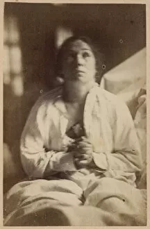
Background imageNeurology Collection: Hysteria, normal appearance, illustration from Iconographie Photographique de La

Hysteria, normal appearance, illustration from Iconographie Photographique de La
REV287038 Hysteria, normal appearance, illustration from Iconographie Photographique de La Salpetriere by Bourneville et P

Background imageNeurology Collection: Physiognomical studies, from Mecanisme de la Physionomie humaine ou analyse

Physiognomical studies, from Mecanisme de la Physionomie humaine ou analyse
CHT207806 Physiognomical studies, from Mecanisme de la Physionomie humaine ou analyse electrophysiologique des passions by Duchenne de Boulogne, published 1876

Background imageNeurology Collection: Physiognomical studies, from 22 to 28, from Mecanisme de la Physionomie humaine ou

Physiognomical studies, from 22 to 28, from Mecanisme de la Physionomie humaine ou
CHT207798 Physiognomical studies, from 22 to 28, from Mecanisme de la Physionomie humaine ou analyse electrophysiologique des passions by Duchenne de Boulogne, published 1876

Background imageNeurology Collection: Hysterical epilepsy, attack, contortions, fig. 2 from

Hysterical epilepsy, attack, contortions, fig. 2 from
REV287076 Hysterical epilepsy, attack, contortions, fig. 2 from Iconographie Photographique de La Salpetriere by Bourneville et P

Background imageNeurology Collection: Hysterical epilepsy, attack, contortions, fig. 1 from

Hysterical epilepsy, attack, contortions, fig. 1 from
REV287075 Hysterical epilepsy, attack, contortions, fig. 1 from Iconographie Photographique de La Salpetriere by Bourneville et P

Background imageNeurology Collection: Hysterical epilepsy, attack, terminal phase, ecstasy, illustration from

Hysterical epilepsy, attack, terminal phase, ecstasy, illustration from
REV287054 Hysterical epilepsy, attack, terminal phase, ecstasy, illustration from Iconographie Photographique de La Salpetriere by Bourneville et P

Background imageNeurology Collection: Hysterical epilepsy, terminal phase, melancholic delirium

Hysterical epilepsy, terminal phase, melancholic delirium
REV287052 Hysterical epilepsy, terminal phase, melancholic delirium, illustration from Iconographie Photographique de La Salpetriere by Bourneville et P

Background imageNeurology Collection: Dementia, General Paralysis, illustration from Nouveau traite elementaire et

Dementia, General Paralysis, illustration from Nouveau traite elementaire et
CHT213235 Dementia, General Paralysis, illustration from Nouveau traite elementaire et pratique des maladies mentales by Henri Dagonet (1823-1902) Paris, Bailliere

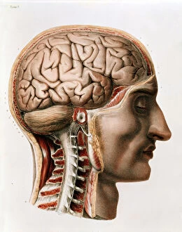
Background imageNeurology Collection: The Brain, plate from Traite Complet de l Anatomie de l Homme

The Brain, plate from Traite Complet de l Anatomie de l Homme
CHT234696 The Brain, plate from Traite Complet de l Anatomie de l Homme by Jean-Baptiste Marc Bourgery (1797-1849) 1866-67 (coloured engraving) by Jacob

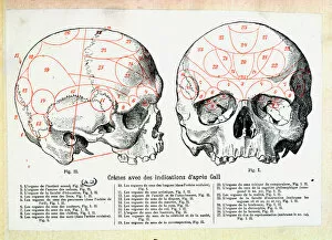
Background imageNeurology Collection: Skull with phrenologic indications after Franz Josef Gall (1758-1828) (engraving)

Skull with phrenologic indications after Franz Josef Gall (1758-1828) (engraving)
CHT164123 Skull with phrenologic indications after Franz Josef Gall (1758-1828) (engraving) by French School, (19th century); Bibliotheque des Arts Decoratifs, Paris

Background imageNeurology Collection: Newcastle General Hospital. Neuro Unit. Circa 1964

Newcastle General Hospital. Neuro Unit. Circa 1964

Background imageNeurology Collection: Brain activity, artwork

Brain activity, artwork
Brain activity, computer artwork

Background imageNeurology Collection: Exterior of National Hospital for the Paralysed and Epileptic, Queen Square, Bloomsbury

Exterior of National Hospital for the Paralysed and Epileptic, Queen Square, Bloomsbury, London. Date: 1885

Background imageNeurology Collection: Figure 26: Expression of painful attention and attention, attentive gaze

Figure 26: Expression of painful attention and attention, attentive gaze. 1854-56, printed 1862

Background imageNeurology Collection: Figure 53: Whimpering and false laughter, 1854-56, printed 1862

Figure 53: Whimpering and false laughter, 1854-56, printed 1862

Background imageNeurology Collection: Figure 39: The attention attracted by an object that provokes lascivious

Figure 39: The attention attracted by an object that provokes lascivious ideas and desires. 1854-56, printed 1862

Background imageNeurology Collection: Figure 9: A study of m. frontalis in maximum contraction, 1854-56, printed 1862

Figure 9: A study of m. frontalis in maximum contraction, 1854-56, printed 1862

Background imageNeurology Collection: Figure 15: Meditation, mental concentration, 1854-56, printed 1862

Figure 15: Meditation, mental concentration, 1854-56, printed 1862

Background imageNeurology Collection: Figure 27: Expression proportionally more pained, 1854-56, printed 1862

Figure 27: Expression proportionally more pained, 1854-56, printed 1862

Background imageNeurology Collection: Figure 81: Lady Macbeth, moderate expression of cruelty, 1854-56, printed 1862

Figure 81: Lady Macbeth, moderate expression of cruelty, 1854-56, printed 1862

Background imageNeurology Collection: Figure 42: Gaiety expressed by the ideas of lustfulness, cynicism, and le

Figure 42: Gaiety expressed by the ideas of lustfulness, cynicism, and lewdness. 1854-56, printed 1862


All Professionally Made to Order for Quick Shipping

Neurology, the fascinating study of the nervous system, unravels the intricate workings of our brain and its various components. From the Motor homunculus model that depicts how different body parts are represented in our motor cortex to the Histological Diagram of a Mammalian Retina revealing the complexity of our visual processing, every aspect is awe-inspiring. The Cerebellum tissue, as seen through a light micrograph, showcases its unique structure responsible for coordinating movement and balance. Meanwhile, an inferior view of the Anatomy of human brain highlights its remarkable organization and interconnectedness. Through Brain fibres captured by DTI MRI scans like C017 / 7099 or C017 / 7035, we gain insights into neural pathways that facilitate communication between different regions. These pathways form a complex network crucial for transmitting information throughout our brain. Exploring Brain blood vessels using a 3D angiogram (C007 / 1981) reveals their vital role in supplying oxygen and nutrients to sustain neuronal function. Additionally, understanding Motor and sensory homunculi helps us comprehend how specific areas within our brains control different bodily functions. Delving deeper into neurology brings us face-to-face with Medulla oblongata artwork depicting this critical region involved in regulating essential autonomic functions such as breathing and heart rate. Furthermore, observing Nerve and glial cells under a light microscope illustrates their diverse roles in supporting neurons' health and functionality. Finally, examining Synapse nerve junctions through TEM unveils these microscopic structures where electrical signals are transmitted from one neuron to another—a fundamental process underlying all brain activity. In essence, neurology takes us on an incredible journey deep within ourselves—unveiling mysteries at both macroscopic levels like anatomical structures or microscopically exploring cellular intricacies, and is through this exploration that we gain profound knowledge about how our brains work—the very essence of who we are as individuals.