Fraud Blocker Skip to main content

Neurology Collection (page 2)

Neurology, the fascinating study of the nervous system, unravels the intricate workings of our brain and its various components

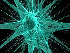
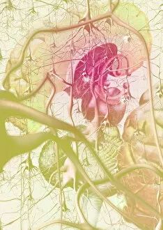
Background imageNeurology Collection: Nerve cells, abstract artwork

Nerve cells, abstract artwork
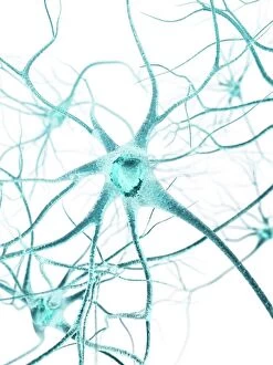
Nerve cells. Abstract computer artwork of nerve cells, or neurons. Neurons are responsible for passing information around the central nervous system (CNS) and from the CNS to the rest of the body

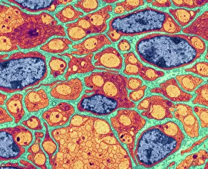
Background imageNeurology Collection: Myelination of nerve fibres, TEM

Myelination of nerve fibres, TEM
Myelination of nerve fibres. Coloured transmission electron micrograph (TEM) of Schwann cells (red, with blue nuclei) insulating nerve fibres (axons, orange) with a myelin sheath
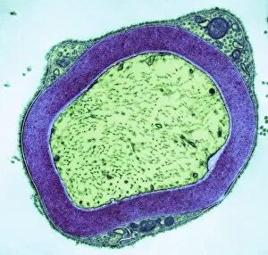
Background imageNeurology Collection: Myelinated nerve, TEM

Myelinated nerve, TEM
Myelinated nerve. Coloured transmission electron micrograph (TEM) of myelinated nerve fibres and Schwann cells. Myelin (purple) is an insulating fatty layer that surrounds nerve fibres (axons)

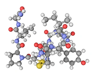
Background imageNeurology Collection: Oxytocin neurotransmitter molecule

Oxytocin neurotransmitter molecule. Computer model showing the structure of the neurotransmitter and hormone Oxytocin. Atoms are colour-coded spheres (carbon: dark grey, hydrogen: light grey)

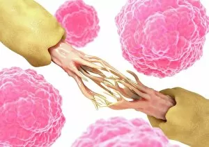
Background imageNeurology Collection: Synapse nerve junctions, SEM

Synapse nerve junctions, SEM
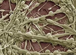
Synapse nerve junctions. Coloured scanning electron micrograph (SEM) of nerve cells showing the synapses (junctions, bulges) between them

Background imageNeurology Collection: White matter fibres, DTI scan

White matter fibres, DTI scan
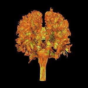
White matter fibres. Coloured 3D diffusion tensor imaging (DTI) scan of the brain, showing the bundles of white matter nerve fibres

Background imageNeurology Collection: French neurologist Duchenne de Boulogne using his electrophysiology apparatus on an old man

French neurologist Duchenne de Boulogne using his electrophysiology apparatus on an old man, 1862 (b / w photo)
6014967 French neurologist Duchenne de Boulogne using his electrophysiology apparatus on an old man, 1862 (b/w photo) by Duchenne de Boulogne

Background imageNeurology Collection: The science of human anatomy by Bartholomeo Eustachi

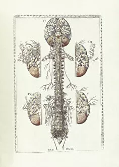
The science of human anatomy by Bartholomeo Eustachi, depicting the shape, size and relative position of the organs of the human body. Circa 18th century

Background imageNeurology Collection: Nerve cell, artwork F007 / 7448

Nerve cell, artwork F007 / 7448
Nerve cell, computer artwork

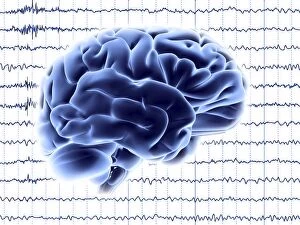
Background imageNeurology Collection: Brain activity, artwork F006 / 4594

Brain activity, artwork F006 / 4594
Brain activity. Computer artwork of EEG (electroencephalogram) traces and a brain illustration. An EEG records the brains activity

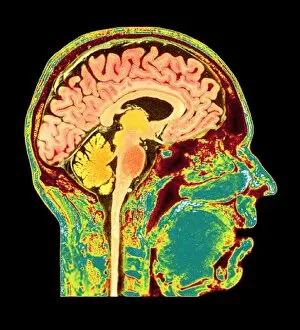
Background imageNeurology Collection: Normal human brain, MRI scan C016 / 8850

Normal human brain, MRI scan C016 / 8850
Brain. Coloured magnetic resonance imaging (MRI) scan of a sagittal section through a patients head showing a healthy human brain

Background imageNeurology Collection: Purkinje nerve cell, TEM C014 / 0583

Purkinje nerve cell, TEM C014 / 0583
Purkinje nerve cell. Transmission electron micrograph (TEM) of a purkinje nerve cell (green) from the cerebellum of the brain, showing the cell body (centre) and its primary dendrite (cell process)

Background imageNeurology Collection: Brain anatomy

Brain anatomy. Computer artwork of strands of nerve cells (neurons) in front of an exploded view of the brain seen from behind

Background imageNeurology Collection: Brain

Brain. Computer artwork of a human brain inside the skull

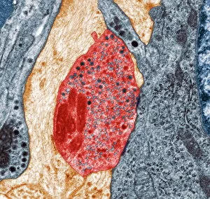
Background imageNeurology Collection: Nerve synapse, TEM

Nerve synapse, TEM
Nerve synapse. Coloured transmission electron micrograph (TEM) of the neuron (nerve) terminal at a synapse in the diaphragm

Background imageNeurology Collection: Nerve cell, SEM

Nerve cell, SEM
Nerve cell. Coloured scanning electron micrograph (SEM) of a neuron (nerve cell). The cell body is the central structure with neurites (long and thin structures) radiating outwards from it

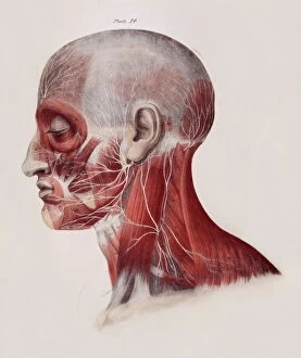
Background imageNeurology Collection: Facial nerves

Facial nerves. Historical anatomical artwork of a side view of a dissected human head showing the nerves (white) and muscles (red) of the face and neck

Background imageNeurology Collection: Nerve cells, computer artwork

Nerve cells, computer artwork
Nerve cells. Computer artwork of nerve cells, or neurons

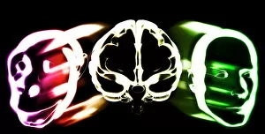
Background imageNeurology Collection: Primate brain evolution

Primate brain evolution. Conceptual computer artwork of a brain (centre) with a monkey head (left) and a human head (right), representing the evolution of primate brains

Background imageNeurology Collection: Nerve damage and stem cells, artwork

Nerve damage and stem cells, artwork
Nerve damage and stem cells, computer artwork. Stem cells are undifferentiated cells that can produce other types of cell when they divide

Background imageNeurology Collection: Slipped intervertebral disc, artwork

Slipped intervertebral disc, artwork
Slipped intervertebral disc, computer artwork. Intervertebral discs (red) are pads of cartilage between the bones (vertebrae) of the spine

Background imageNeurology Collection: Serotonin neurotransmitter molecule

Serotonin neurotransmitter molecule
Serotonin molecule. Computer artwork of a molecule of serotonin (5-hydroxytryptamine, C10.H12.N2.O), a neurotransmitter. Atoms (spheres) are colour- coded: carbon (black), hydrogen (white)

Background imageNeurology Collection: Jean Martin Charcot's electrotherapy clinic

Jean Martin Charcot's electrotherapy clinic
5311887 Jean Martin Charcot's electrotherapy clinic; (add.info.: Engraving depicting Jean Martin Charcot's electrotherapy clinic at the Salpetriere, Paris, founded 1877)

Background imageNeurology Collection: Map of nerve bundles in the human brain by Paul Broca

Map of nerve bundles in the human brain by Paul Broca
5309460 Map of nerve bundles in the human brain by Paul Broca; (add.info.: Map of nerve bundles in the human brain by Paul Broca)

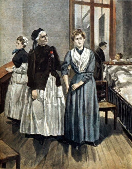
Background imageNeurology Collection: Nurse Marguerite Bottard (1822-1906), at the Paris Psychiatric Hospital. 1841 (watercolour)

Nurse Marguerite Bottard (1822-1906), at the Paris Psychiatric Hospital. 1841 (watercolour)
5669372 Nurse Marguerite Bottard (1822-1906), at the Paris Psychiatric Hospital. 1841 (watercolour); Universal History Archive/UIG

Background imageNeurology Collection: Jean Martin Charcot (1825-1893 French neurologist and hypnotist, engraving, colourized document)

Jean Martin Charcot (1825-1893 French neurologist and hypnotist, engraving, colourized document)
1745971 Jean Martin Charcot (1825-1893 French neurologist and hypnotist, engraving, colourized document; (add.info.: Jean Martin Charcot (1825-1893 French neurologist and hypnotist, engraving)

Background imageNeurology Collection: Jean Martin Charcot (1825-1903) French neurologist and pathologist

Jean Martin Charcot (1825-1903) French neurologist and pathologist, demonstrating production of hypnosis using beam of
542255 Jean Martin Charcot (1825-1903) French neurologist and pathologist, demonstrating production of hypnosis using beam of light from a magic lantern

Background imageNeurology Collection: Jean-Martin Charcot

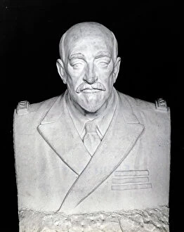
Jean-Martin Charcot
3732975 Jean-Martin Charcot; (add.info.: Bust of Jean-Martin Charcot (1825-1893) a French neurologist and professor of anatomical pathology. Dated 19th Century.); Universal History Archive/UIG

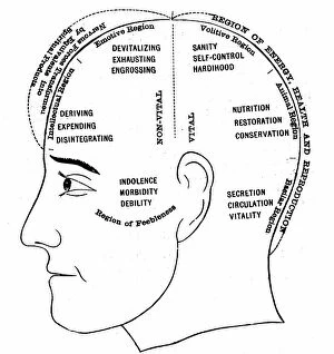
Background imageNeurology Collection: An engraving depicting a diagram showing the supposed functions of various areas of the brain

An engraving depicting a diagram showing the supposed functions of various areas of the brain, 19th century
5311364 An engraving depicting a diagram showing the supposed functions of various areas of the brain, 19th century; (add.info.: Date: 1886); Universal History Archive/UIG

Background imageNeurology Collection: Portrait of Jean Martin Charcot (1825-1893), French physician clinician, neurologist

Portrait of Jean Martin Charcot (1825-1893), French physician clinician, neurologist
ELD4902763 Portrait of Jean Martin Charcot (1825-1893), French physician clinician, neurologist and professor of anatomical pathology

Background imageNeurology Collection: An engraving depicting Descartes drawing of the human brain, 17th century

An engraving depicting Descartes drawing of the human brain, 17th century
5311365 An engraving depicting Descartes drawing of the human brain, 17th century; Universal History Archive/UIG

Background imageNeurology Collection: Jean-Martin Charcot

Jean-Martin Charcot
3078440 Jean-Martin Charcot; (add.info.: Portrait of Jean-Martin Charcot (1825-1893) a French neurologist and professor of anatomical pathology. Dated 19th Century.); Universal History Archive/UIG

Background imageNeurology Collection: William Henry Broadbent (1835-1907)

William Henry Broadbent (1835-1907)
865196 William Henry Broadbent (1835-1907); (add.info.: William Henry Broadbent (1835-1907) English physician born at Lindley, near Huddersfield, Yorkshire

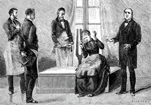
Background imageNeurology Collection: Jean Martin Charcot (1825-1903) French neurologist and pathologist, on right, 1879 (engraving)

Jean Martin Charcot (1825-1903) French neurologist and pathologist, on right, 1879 (engraving)
542252 Jean Martin Charcot (1825-1903) French neurologist and pathologist, on right, 1879 (engraving); (add.info.: demonstrating production of hypnosis using the sound from a large tuning fork)

Background imageNeurology Collection: Jean Martin Charcot (1825-1893) giving a clinical lecture at the Salpetriere, 1887 (engraving)

Jean Martin Charcot (1825-1893) giving a clinical lecture at the Salpetriere, 1887 (engraving)
542287 Jean Martin Charcot (1825-1893) giving a clinical lecture at the Salpetriere, 1887 (engraving); (add.info.: Engraving after painting by Andre Brouillet)

Background imageNeurology Collection: Professor Ivan Petrovich Pavlov, Russian scientist on a visit to London (b/w photo)

Professor Ivan Petrovich Pavlov, Russian scientist on a visit to London (b/w photo)
8664296 Professor Ivan Petrovich Pavlov, Russian scientist on a visit to London (b/w photo) by English Photographer, (20th century); Private Collection; (add.info)

Background imageNeurology Collection: A cartoon commenting on the fashionable revival of phrenology, 19th century

A cartoon commenting on the fashionable revival of phrenology, 19th century
5311366 A cartoon commenting on the fashionable revival of phrenology, 19th century; (add.info.: Date: 1878); Universal History Archive/UIG

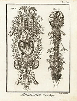
Background imageNeurology Collection: The nervous system. Copperplate engraving by Robert Benard after illustrations by Raymond Vieussens

The nervous system. Copperplate engraving by Robert Benard after illustrations by Raymond Vieussens
FLO4611615 The nervous system. Copperplate engraving by Robert Benard after illustrations by Raymond Vieussens and Bartolomeo Eustachi from Denis Diderot's Encyclopedia, Pellet, Geneva

Background imageNeurology Collection: Portrait of Jean-Martin Charcot

Portrait of Jean-Martin Charcot
673308 Portrait of Jean-Martin Charcot; (add.info.: Jean-Martin Charcot (29 November 1825 - 16 August 1893, French neurologist and professor of anatomical pathology, ); Universal History Archive/UIG

Background imageNeurology Collection: Jean Martin Charcot (1825-1893) French neurologist c. 1880, engraving

Jean Martin Charcot (1825-1893) French neurologist c. 1880, engraving
1750927 Jean Martin Charcot (1825-1893) French neurologist c. 1880, engraving; (add.info.: Jean Martin Charcot (1825-1893) French neurologist c. 1880, engraving); Photo: Tallandier

Background imageNeurology Collection: Professor Jean-Martin Charcot, c.1870 (b/w photo)

Professor Jean-Martin Charcot, c.1870 (b/w photo)
1950024 Professor Jean-Martin Charcot, c.1870 (b/w photo); (add.info.: Jean Martin Charcot (1825-1893) french neurologist c. 1870); Photo © CCI

Background imageNeurology Collection: Phrenological heads, 19th century (object)

Phrenological heads, 19th century (object)
1828274 Phrenological heads, 19th century (object); (add.info.: Heads used to study phrenology, they belong to doctor Jean-Martin Charcot (1825-1893)); Photo © CCI

Background imageNeurology Collection: Hysterical women in the courtyard of the parisian hospital of La salpetriere studied by the French

Hysterical women in the courtyard of the parisian hospital of La salpetriere studied by the French neurologist
ELD4867797 Hysterical women in the courtyard of the parisian hospital of La salpetriere studied by the French neurologist Jean-Martin Charcot (1825-1893), 1885

Background imageNeurology Collection: Jean-Martin Charcot (1825-1893) French neurologist, photo Nadar, 1890

Jean-Martin Charcot (1825-1893) French neurologist, photo Nadar, 1890
1758795 Jean-Martin Charcot (1825-1893) French neurologist, photo Nadar, 1890; (add.info.: Jean-Martin Charcot (1825-1893) French neurologist, photo Nadar, 1890); Photo: Tallandier

Background imageNeurology Collection: Sigrid Hjertén, 1931. Creator: Isaac Grünewald

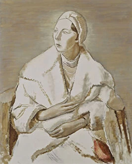
Sigrid Hjertén, 1931. Creator: Isaac Grünewald
Sigrid Hjerten, 1931

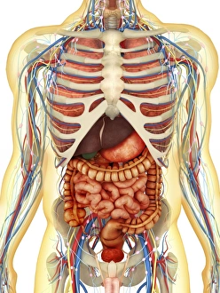
Background imageNeurology Collection: Human body with internal organs, nervous system, lymphatic system and circulatory system

Human body with internal organs, nervous system, lymphatic system and circulatory system
Transparent human body with internal organs, nervous system, lymphatic system and circulatory system

Background imageNeurology Collection: Substantia nigra in Parkinsons disease, illustration

Substantia nigra in Parkinsons disease, illustration
Substantia nigra. Computer illustration showing a degenerated substantia nigra and dopaminergic neurons in Parkinsons disease


All Professionally Made to Order for Quick Shipping

Neurology, the fascinating study of the nervous system, unravels the intricate workings of our brain and its various components. From the Motor homunculus model that depicts how different body parts are represented in our motor cortex to the Histological Diagram of a Mammalian Retina revealing the complexity of our visual processing, every aspect is awe-inspiring. The Cerebellum tissue, as seen through a light micrograph, showcases its unique structure responsible for coordinating movement and balance. Meanwhile, an inferior view of the Anatomy of human brain highlights its remarkable organization and interconnectedness. Through Brain fibres captured by DTI MRI scans like C017 / 7099 or C017 / 7035, we gain insights into neural pathways that facilitate communication between different regions. These pathways form a complex network crucial for transmitting information throughout our brain. Exploring Brain blood vessels using a 3D angiogram (C007 / 1981) reveals their vital role in supplying oxygen and nutrients to sustain neuronal function. Additionally, understanding Motor and sensory homunculi helps us comprehend how specific areas within our brains control different bodily functions. Delving deeper into neurology brings us face-to-face with Medulla oblongata artwork depicting this critical region involved in regulating essential autonomic functions such as breathing and heart rate. Furthermore, observing Nerve and glial cells under a light microscope illustrates their diverse roles in supporting neurons' health and functionality. Finally, examining Synapse nerve junctions through TEM unveils these microscopic structures where electrical signals are transmitted from one neuron to another—a fundamental process underlying all brain activity. In essence, neurology takes us on an incredible journey deep within ourselves—unveiling mysteries at both macroscopic levels like anatomical structures or microscopically exploring cellular intricacies, and is through this exploration that we gain profound knowledge about how our brains work—the very essence of who we are as individuals.