
Naturemycology Collection
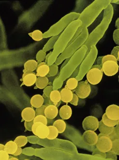
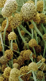
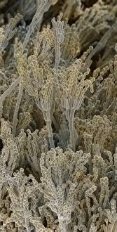
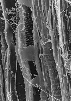
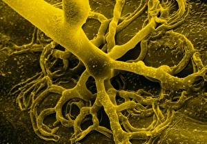
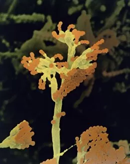
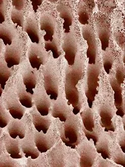
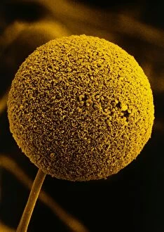
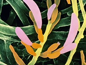
Exploring the intricate world of nature's mycology 🍄✨ From the fascinating SEM images of penicillin fungus


All Professionally Made to Order for Quick Shipping
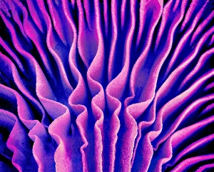
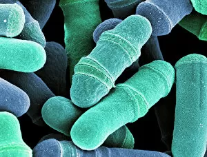
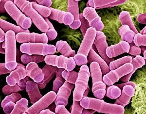
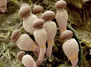
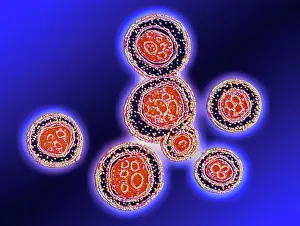
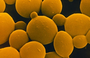
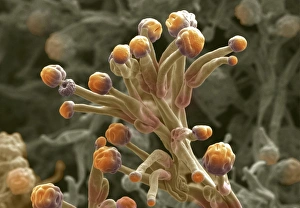
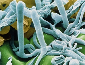
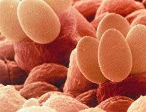
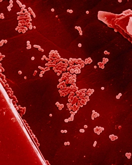
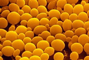
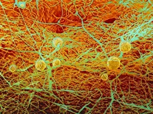
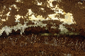
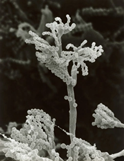
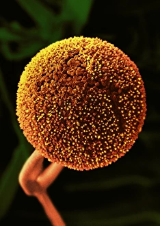
Exploring the intricate world of nature's mycology 🍄✨ From the fascinating SEM images of penicillin fungus and mushroom gills to the mesmerizing art of yeast cell budding, this captivating journey takes us deep into the microscopic wonders. Witness the beauty of Cep mushrooms (Boletus edulis) and their intricate structures under SEM, while marveling at dividing yeast cells as they multiply in a symphony of life. Delve into LM imagery revealing the fruiting bodies of Rhizopus oligosporus and Cryptococcus neoformans fungi, each with its unique characteristics. Candida albicans yeast showcases its delicate form through stunning SEM visuals, reminding us that even tiny organisms can hold immense beauty. Explore Penicillium fungal spores in breathtaking detail under SEM, uncovering their remarkable patterns and textures. Lastly, get acquainted with Pilobolus fungus - an intriguing species that never fails to amaze with its distinctive appearance. Naturemycology invites you to immerse yourself in these extraordinary glimpses into nature's hidden kingdom.