Fraud Blocker Skip to main content

Ligaments Collection (page 4)

"Ligaments: The Unsung Heroes of Our Body's Stability and Mobility" Damaged knee ligament, artwork: A visual representation of the intricate network in our knees

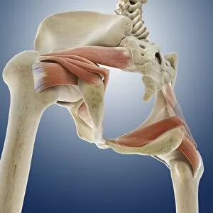
Background imageLigaments Collection: Buttock muscles, artwork C013 / 4420

Buttock muscles, artwork C013 / 4420
Buttock muscles. Computer artwork of a posterior view of the hips showing some of the muscles (red) of the buttocks. These are shown attached to the femurs (thigh bones) and the pelvis bones

Background imageLigaments Collection: Buttock muscles, artwork C013 / 4421

Buttock muscles, artwork C013 / 4421
Buttock muscles. Computer artwork of a side view (front of body at left) of the hips showing some of the muscles (red) of the buttocks

Background imageLigaments Collection: Buttock muscles, artwork C013 / 4419

Buttock muscles, artwork C013 / 4419
Buttock muscles. Computer artwork of a posterior oblique view of the hips showing some of the muscles (red) of the buttocks. These are shown attached to the femurs (thigh bones) and the pelvis bones

Background imageLigaments Collection: Buttock muscles, artwork C013 / 4415

Buttock muscles, artwork C013 / 4415
Buttock muscles. Computer artwork of a posterior oblique view of the hips showing some of the muscles (red) of the buttocks. These are shown attached to the femurs (thigh bones) and the pelvis bones

Background imageLigaments Collection: Buttock muscles, artwork C013 / 4416

Buttock muscles, artwork C013 / 4416
Buttock muscles. Computer artwork of a posterior oblique view of the hips showing some of the muscles (red) of the buttocks. These are shown attached to the femurs (thigh bones) and the pelvis bones

Background imageLigaments Collection: Muscles of the hand

Muscles of the hand

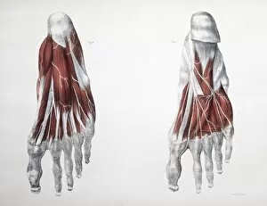
Background imageLigaments Collection: Muscles of the foot

Muscles of the foot, historical artwork. The figure at top left shows the first layer of muscles (red) in the sole of the foot. The skin and fascia (connective tissue) have been removed

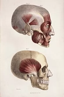
Background imageLigaments Collection: Head muscles

Head muscles
Head musculature. Historical artwork of the facial and other head muscles (red) on a human skull. The temporal muscle is shown in the lower frame

Background imageLigaments Collection: Muscles of the back

Muscles of the back, historical artwork. The skin, fascia (connective tissue), and first three layers of muscles have been removed to expose the fourth muscle layer (red) of the back

Background imageLigaments Collection: Hand anatomy

Hand anatomy, computer artwork. Muscles are pink, tendons, ligaments and bones are cream. Tendons and ligaments are bands of inelastic fibrous tissue

Background imageLigaments Collection: Foot nerves

Foot nerves. Historical anatomical artwork of the nerves (white) of the sole of a human foot. Also shown are muscles (red), and ligaments, tendons and other connective tissues (grey)

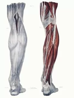
Background imageLigaments Collection: Leg nerves

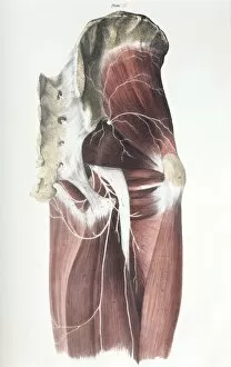
Leg nerves. Historical anatomical artwork of the nerves (white) of the rear of a human lower leg. At left, the superficial (surface) nerves are seen and at right the deep nerves and muscles (red)

Background imageLigaments Collection: Pelvic spinal nerves

Pelvic spinal nerves. Historical anatomical artwork of the spinal nerves (white) in the human pelvic region. This side view (front of the body at right)

Background imageLigaments Collection: Knee bones and ligaments

Knee bones and ligaments. Historical anatomical artwork of knee bones (yellow) and ligaments (pale blue). Ligaments are bands of fibrous tissue that hold bones together at joints

Background imageLigaments Collection: Hand bones and ligaments

Hand bones and ligaments. Historical anatomical artwork of the bones (yellow) & ligaments (white) of the hand. Ligaments are bands of fibrous tissue that hold bones together at their joints

Background imageLigaments Collection: Lower arm bones and ligaments

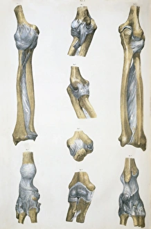
Lower arm bones and ligaments
Lower arm bones & ligaments. Historical anatomical artwork of lower arm bones (yellow) and ligaments (pale blue). Ligaments are bands of fibrous tissue that hold bones together at joints

Background imageLigaments Collection: Ankle bones and ligaments

Ankle bones and ligaments. Historical anatomical artwork of ankle bones (yellow) and ligaments (pale blue). Ligaments are bands of fibrous tissue that hold bones together at their joints

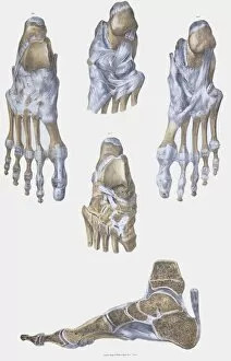
Background imageLigaments Collection: Foot bones and ligaments

Foot bones and ligaments. Historical anatomical artwork of foot bones (yellow) and ligaments (pale blue). Ligaments are bands of fibrous tissue that hold bones together at their joints

Background imageLigaments Collection: Lower leg bones and ligaments

Lower leg bones and ligaments. Historical anatomical artwork of lower leg bones (yellow) and ligaments (pale blue). Ligaments are bands of fibrous tissue that hold bones together at their joints

Background imageLigaments Collection: Pelvis bones and ligaments

Pelvis bones and ligaments. Historical anatomical artwork of pelvis bones (yellow) and ligaments (white). Ligaments are bands of fibrous tissue that hold bones together at their joints

Background imageLigaments Collection: Shoulder bones and ligaments

Shoulder bones and ligaments. Historical anatomical artwork of shoulder bones (yellow) and ligaments (white). Ligaments are bands of fibrous tissue that hold bones together at their joints

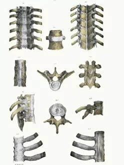
Background imageLigaments Collection: Spinal bones and ligaments

Spinal bones and ligaments. Historical anatomical artwork of thoracic (upper) spinal bones (yellow, vertebrae) and ligaments (white)

Background imageLigaments Collection: Apleys knee test

Apleys knee test
MODEL RELEASED. Apleys knee test. Physiotherapist performing an Apleys knee test to assess a patients knee injury. This test is carried out to diagnose knee cartilage injuries

Background imageLigaments Collection: Lachman knee ligament test

Lachman knee ligament test
MODEL RELEASED. Lachman knee ligament test. Physiotherapist performing a Lachman test on a patients knee ligaments. This test is designed as an assessment of the knees anterior cruciate ligament

Background imageLigaments Collection: Eye anatomy, SEM

Eye anatomy, SEM
Eye anatomy. Coloured scanning electron micrograph (SEM) showing part of the ciliary body (blue) and iris (right) of an eye

Background imageLigaments Collection: Eye lens and accommodation, diagram

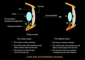
Eye lens and accommodation, diagram
Eye lens and accommodation. Diagram showing the process by which the eye changes the shape of the lens when changing between close vision and distant vision. This process is known as accommodation

Background imageLigaments Collection: Knee in anterior view, artwork

Knee in anterior view, artwork
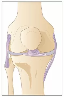
Artwork of the anatomy of the knee joint (anterior view) showing the femur (thigh bone, at top) articulating with the tibia (shin bone, at bottom), cartilage (light blue)

Background imageLigaments Collection: Knee joint anatomy, artwork

Knee joint anatomy, artwork
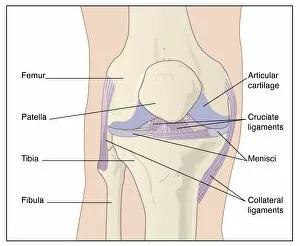
Artwork of the anatomy of the knee joint (anterior view) showing the femur (thigh bone, at top) articulating with the tibia (shin bone, at bottom), articular cartilage

Background imageLigaments Collection: Cruciate ligament injury, artwork

Cruciate ligament injury, artwork
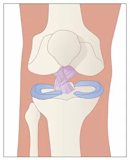
Artwork (anterior view) of the anatomy of the knee joint showing the inner cruciate ligaments (purple) arranged like a cross which serve to stabilize the knee joint while allowing motion

Background imageLigaments Collection: Eye lens and accommodation, artwork

Eye lens and accommodation, artwork
Eye lens and accommodation. Artwork showing the process by which the eye changes the shape of the lens when changing between close vision and distant vision. This process is known as accommodation

Background imageLigaments Collection: Anterior cruciate ligament tear, CT scan

Anterior cruciate ligament tear, CT scan
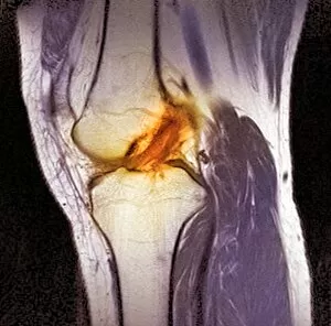
Anterior cruciate ligament tear. Coloured computer tomography (CT) scan of the knee joint of a patient with a tear of the anterior cruciate ligament (ACL)

Background imageLigaments Collection: Normal human knee, artwork

Normal human knee, artwork
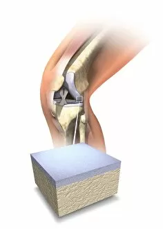
Normal human knee. Artwork showing the internal anatomy of a healthy human knee, with an inset (bottom) of the cartilage (grey) that covers the ends of the bones forming this joint

Background imageLigaments Collection: Damaged knee, artwork

Damaged knee, artwork
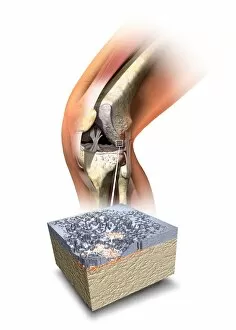
Damaged knee. Artwork showing the internal anatomy of a damaged human knee, with an inset (bottom) of the cartilage (grey) that covers the ends of the bones forming this joint

Background imageLigaments Collection: Wrist Brace

Wrist Brace
Wrist sprain injuries caused by damage to the ligaments can be helped by this form of adjustable wrist brace made from woven elastic and having a metal stay incorporated in the palm area

Background imageLigaments Collection: Skeleton and ligaments, artwork

Skeleton and ligaments, artwork
Female skeleton, computer artwork. The human skeleton has 206 bones. The skull (at top) protects the brain. The ribs of the chest (at upper centre) enclose the heart and lungs


All Professionally Made to Order for Quick Shipping

"Ligaments: The Unsung Heroes of Our Body's Stability and Mobility" Damaged knee ligament, artwork: A visual representation of the intricate network in our knees, highlighting the importance of their role in supporting joint stability. Skeleton & Ligaments: Explore the hidden framework beneath our skin as we delve into the fascinating connection between our skeleton and ligaments that enable us to move with grace. Arm circulation, anatomical artwork C013 / 7419: Discover how ligaments work hand-in-hand with blood vessels to ensure proper arm circulation, allowing us to perform everyday tasks effortlessly. Muscles of the neck: Unveil the interplay between muscles and ligaments in our neck region, crucial for maintaining posture and facilitating smooth head movements. Eye muscle, TEM C014 / 1468: Delicate yet powerful eye muscles intricately intertwined with resilient ligaments—uncover this captivating combination responsible for precise eye movements. Outer ankle ligaments, artwork C013 / 4452: Step into a world where outer ankle ligaments provide stability during every stride we take—a testament to their resilience against external forces. Inner ankle ligaments, artwork C013 / 4451: Journey deeper into understanding inner ankle ligament anatomy as they safeguard against inward rolling motions while walking or running. Eye anatomy, SEM: Peer through a scanning electron microscope lens at detailed images revealing how tiny but mighty eye structures rely on robustly connected networks of supportive ligaments. Muscles (engraving): Witness an enchanting engraving showcasing various muscles working harmoniously alongside essential connective tissues like tendons and—of course—ligaments. Ligaments of the human body (engraving): An artistic masterpiece capturing an array of interconnected human body parts held together by strong bands known as "ligaments"—a true testament to the body's architectural brilliance.