Home > Popular Themes > Human Body
Head muscles
![]()

Wall Art and Photo Gifts from Science Photo Library
Head muscles
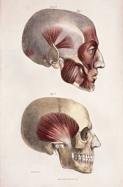
Head musculature. Historical artwork of the facial and other head muscles (red) on a human skull. The temporal muscle is shown in the lower frame. The upper frame shows the muscles attached to the ear, which are not very developed in humans. The facial muscles are well developed, allowing the extensive facial expressions (with the eyes, nose, lips, cheeks and forehead) that are an important part of non-verbal communication. The large and powerful jaw muscles are also seen. Published in The Muscles of the Human Body by Jones Quain in 1836
Science Photo Library features Science and Medical images including photos and illustrations
Media ID 6448455
© SHEILA TERRY/SCIENCE PHOTO LIBRARY
1836 19th Bones Communication Deep Expression Eyes Face Facial Forehead Historical Image Imagery Language Layered Ligament Ligaments Lips Lower Mandible Mouth Muscle System Muscles Muscular Nineteenth Century Nose Ocular Profile Side Superficial Temple Temporal Tendon Tendons Jones Musculature Quain
EDITORS COMMENTS
This historical artwork showcases the intricate and layered musculature of the human head. Published in 1836 by Jones Quain, it provides a detailed glimpse into the complex structure responsible for our facial expressions and non-verbal communication. The red-colored muscles depicted on a human skull highlight the well-developed facial muscles that enable us to convey a wide range of emotions through our eyes, nose, lips, cheeks, and forehead. These expressive features play an essential role in conveying messages without words. In this fascinating illustration, attention is drawn to the temporal muscle showcased in the lower frame. Its prominence emphasizes its power and significance in facilitating jaw movement. The upper frame focuses on less developed ear muscles found in humans compared to other species. Not only does this image offer valuable insights into anatomical structures but also serves as a testament to scientific advancements during the 19th century. It beautifully captures both superficial and deep layers of head musculature while highlighting key elements such as bones, tendons, ligaments, and ocular connections. As we delve into this historical imagery from Science Photo Library's collection titled "The Muscles of the Human Body" we are reminded of how intricately designed our bodies are. This print invites us to appreciate not only their functional aspects but also their artistic beauty within medical illustrations from centuries past.
MADE IN THE UK
Safe Shipping with 30 Day Money Back Guarantee
FREE PERSONALISATION*
We are proud to offer a range of customisation features including Personalised Captions, Color Filters and Picture Zoom Tools
SECURE PAYMENTS
We happily accept a wide range of payment options so you can pay for the things you need in the way that is most convenient for you
* Options may vary by product and licensing agreement. Zoomed Pictures can be adjusted in the Basket.

