Fraud Blocker Skip to main content

Lateral Collection (page 9)

"Lateral: Exploring the Intricacies of Nature's Design" Delving into the intricate network of brain blood vessels, as captured in a mesmerizing 3D angiogram C007 / 1981

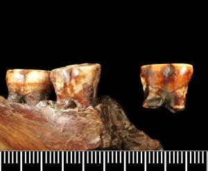
Background imageLateral Collection: Piltdown 1 jaw with Piltdown 2 molar

Piltdown 1 jaw with Piltdown 2 molar

Background imageLateral Collection: Homo sapiens cranium (Florisbad 1)

Homo sapiens cranium (Florisbad 1)
Lateral and frontal view of a cast of a Homo sapiens skull from Florisbad, South Africa. Upper Pleistocene 130, 000-12, 000 BP. Reconstructed by P. Cohen

Background imageLateral Collection: Felis silvestris, cat

Felis silvestris, cat
Photograph of the left view of a domestic cat skull from the education collection at the Natural History Museum at Tring, part of the Natural History Museum, London

Background imageLateral Collection: Oryctolagus cuniculus, European rabbit

Oryctolagus cuniculus, European rabbit
Photograph of the left side view of a European rabbit skull from the education collection at the Natural History Museum at Tring, part of the Natural History Museum, London

Background imageLateral Collection: Dendroaspis angusticeps, green mamba

Dendroaspis angusticeps, green mamba
Photograph of the skull of a green mamba, showing the snakes upper jaw fangs

Background imageLateral Collection: Roman Theatre in Merida. 16 -15 BC. SPAIN. EXTREMADURA

Roman Theatre in Merida. 16 -15 BC. SPAIN. EXTREMADURA

Background imageLateral Collection: Lluza or Llucanes, Master of (begin 13th century)

Lluza or Llucanes, Master of (begin 13th century)
Llua or Llucanes, Master of (begin 13th century). Altar of Santa Maria of Llu.. Detail of a lateral. Scene

Background imageLateral Collection: Silver Birch (Betula pendula) lateral growth of fallen trunk

Silver Birch (Betula pendula) lateral growth of fallen trunk, in snow covered woodland habitat at edge of river valley fen, Redgrave and Lopham Fen N.N.R. Waveney Valley, Suffolk, England, november

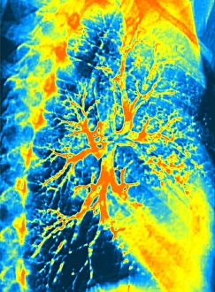
Background imageLateral Collection: Lung bronchogram, coloured X-ray

Lung bronchogram, coloured X-ray
Healthy lung. Coloured bronchogram x-ray of a side (lateral) view of a healthy human lung. The vertebrae of the spinal column are seen at left

Background imageLateral Collection: Ear anatomy

Ear anatomy. Historical anatomical artwork of a human ear. The main diagram (centre) shows the outer ear (pinna, left) and the internal structure of the ear (moving left-right): the ear canal

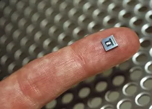
Background imageLateral Collection: Micromechanical accelerometer

Micromechanical accelerometer. This tiny acceleration sensor is used in automatic car stability control systems. These systems can sense lateral (sideways)

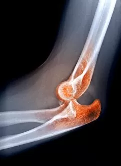
Background imageLateral Collection: Dislocated elbow, X-ray

Dislocated elbow, X-ray
Dislocated elbow. Coloured lateral X-ray of an injury to the elbow (centre) showing that the upper arm bone (humerus, top right) is misaligned (dislocated, turquoise)

Background imageLateral Collection: Knee meniscus tear

Knee meniscus tear
MODEL RELEASED. Knee meniscus tear. Coloured magnetic resonance imaging (MRI) scan of a coronal (front) section through the right knee joint of a 52 year old woman

Background imageLateral Collection: Two views of the human spine

Two views of the human spine. This computer artwork shows a side (lateral) view (left) and an front (anterior) view (right)

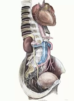
Background imageLateral Collection: Sympathetic nerves, lower body

Sympathetic nerves, lower body
Sympathetic nerves in the lower body. Historical anatomical artwork of a side view of a dissected human abdomen and chest, showing the origin and route of parts of the sympathetic nervous system

Background imageLateral Collection: Sympathetic nerves, upper body

Sympathetic nerves, upper body
Sympathetic nerves in the upper body. Historical anatomical artwork of a side view of a dissected human face, neck and chest, showing the origin and route of parts of the sympathetic nervous system

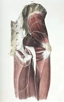
Background imageLateral Collection: Pelvic spinal nerves

Pelvic spinal nerves. Historical anatomical artwork of the spinal nerves (white) in the human pelvic region. This side view (front of the body at right)

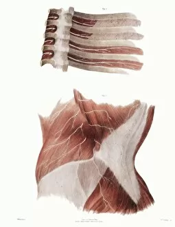
Background imageLateral Collection: Torso nerves

Torso nerves. Historical anatomical artwork of the nerves (white) of parts of a human torso. At lower frame, a side view of the abdomen (front of body at left) shows muscles (red)

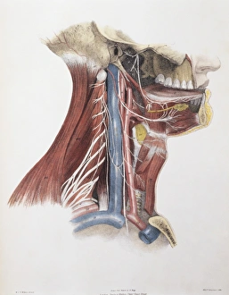
Background imageLateral Collection: Twelfth cranial nerve

Twelfth cranial nerve (hypoglossal nerve, cranial nerve XII). Historical anatomical artwork of a side view of a dissected human neck showing veins (blue), arteries (red), muscles (red)

Background imageLateral Collection: Knee bones and ligaments

Knee bones and ligaments. Historical anatomical artwork of knee bones (yellow) and ligaments (pale blue). Ligaments are bands of fibrous tissue that hold bones together at joints

Background imageLateral Collection: Ankle bones and ligaments

Ankle bones and ligaments. Historical anatomical artwork of ankle bones (yellow) and ligaments (pale blue). Ligaments are bands of fibrous tissue that hold bones together at their joints

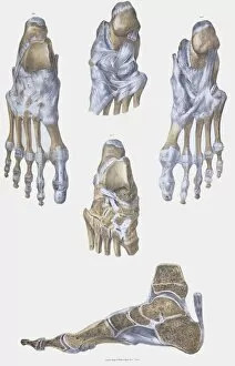
Background imageLateral Collection: Foot bones and ligaments

Foot bones and ligaments. Historical anatomical artwork of foot bones (yellow) and ligaments (pale blue). Ligaments are bands of fibrous tissue that hold bones together at their joints

Background imageLateral Collection: Pelvis bones and ligaments

Pelvis bones and ligaments. Historical anatomical artwork of pelvis bones (yellow) and ligaments (white). Ligaments are bands of fibrous tissue that hold bones together at their joints

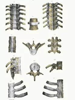
Background imageLateral Collection: Spinal bones and ligaments

Spinal bones and ligaments. Historical anatomical artwork of thoracic (upper) spinal bones (yellow, vertebrae) and ligaments (white)

Background imageLateral Collection: Normal ankle joint, X-ray

Normal ankle joint, X-ray
Normal ankle (talocrural) joint, coloured profile X-ray. The front of the foot is at right, with the heel at lower centre

Background imageLateral Collection: Lumbar spine and sacrum, computer artwork

Lumbar spine and sacrum, computer artwork
Lumbar spine and sacrum, side view, computer artwork. The bones of the spine (vertebrae) are blue and the intervertebral discs are red. The sacrum is at bottom

Background imageLateral Collection: Lumbar spine, computer artwork

Lumbar spine, computer artwork
Lumbar spine, side view, computer artwork. The bones of the spine (vertebrae) are blue and the intervertebral discs are red

Background imageLateral Collection: Anatomical planes of the body, artwork

Anatomical planes of the body, artwork. The three planes shown here are the axial, saggital and coronal. The axial, or transverse, plane splits the body along a horizontal axis

Background imageLateral Collection: Human skeleton, lateral view

Human skeleton, lateral view
This woodcut illustration comes from Chirugia magna by Andreas Vesalius, printed at Venice in 1569

Background imageLateral Collection: Eye anatomy

Eye anatomy. Cutaway artwork passing vertically through a human eye, showing its internal anatomy and structure. The front of the eye is at left, and the structures here include the cornea

Background imageLateral Collection: Head of Pollack Pollachius pollachius a member of the cod family Aquariumgalicia Reboredo Galicia

Head of Pollack Pollachius pollachius a member of the cod family Aquariumgalicia Reboredo Galicia Spain
MAB-294 Head of Pollack, a member of the cod family Aquariumgalicia Reboredo Galicia, Spain Pollachius pollachius Can sometimes be known as Colin Mark Boulton Please note that prints are for personal

Background imageLateral Collection: Pollack Pollachius pollachius a member of the cod family - Aquariumgalicia Reboredo Galicia Spain

Pollack Pollachius pollachius a member of the cod family - Aquariumgalicia Reboredo Galicia Spain
MAB-296 Pollack - a member of the cod family Aquariumgalicia Reboredo Galicia, Spain Pollachius pollachius Can sometimes be known as Colin Mark Boulton Please note that prints are for personal

Background imageLateral Collection: Cayleys Helicopter

Cayleys Helicopter
CAYLEYs HELICOPTER-AEROPLANE With two lateral helicopter rotors to provide lift, and two propellers for propulsion, this is an astonishingly far- sighted proposal


All Professionally Made to Order for Quick Shipping

"Lateral: Exploring the Intricacies of Nature's Design" Delving into the intricate network of brain blood vessels, as captured in a mesmerizing 3D angiogram C007 / 1981. Unveiling the hidden wonders of the human knee joint through an anatomy study that reveals its lateral complexities. Witnessing the majestic Eurasian lynx (Lynx lynx) in all its glory, with its captivating lateral movements and stealthy demeanor. Marveling at the delicate intricacy of brain blood vessels, showcasing nature's remarkable lateral pathways within our most vital organ. Transporting ourselves back to 1956 French Grand Prix, where mechanics examine Trintignants Bugatti Type 251 chassis and its straight 8 Bugatti 251 engine from a lateral perspective. Glimpsing at the striking Red-billed Chough (Pyrrhocorax pyrrhocorax) at Alpine Zoo Innsbruck, Tyrol, Austria, Europe - a testament to nature's lateral adaptations for survival in high altitudes. Admiring an exquisite artwork from 1825 depicting male groin arteries - a fascinating exploration into our body's intricate vascular system from a unique lateral viewpoint. Discovering head and neck muscles through an extraordinary artwork dating back to 1831 - offering insights into their complex interplay and role in our daily movements from various angles including laterally. Examining a tonguestone (sharks tooth) adorned with intriguing lateral denticles that enhance these creatures' feeding efficiency and survival strategies over millions of years. Peering into skull anatomy to unravel mysteries hidden beneath our skin; exploring bone structures and cranial features from both frontal and lateral perspectives for comprehensive understanding. Venturing deep within Earth's core as we explore "Earth's Central Fire, " uncovering geological forces that shape our planet's lateral movements and tectonic shifts.