Fraud Blocker Skip to main content

Lateral Collection (page 6)

"Lateral: Exploring the Intricacies of Nature's Design" Delving into the intricate network of brain blood vessels, as captured in a mesmerizing 3D angiogram C007 / 1981

Background imageLateral Collection: Lateral Elevation of the Thyphonium at Dendera. : lithograph after watercolour

Lateral Elevation of the Thyphonium at Dendera. : lithograph after watercolour
Lateral Elevation of the Thyphonium at Dendera.: lithograph after watercolour by David Roberts (1796-1864) Scottish artist. Ancient Egypt Archaeology

Background imageLateral Collection: Scoliosis of the spine, X-ray C017 / 7167

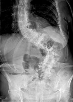
Scoliosis of the spine, X-ray C017 / 7167
Scoliosis. X-ray of the spine of a 71 year old female patient with severe scoliosis (sideways curvature). This condition usually starts in infancy or adolescence, and is most often seen in girls
Background imageLateral Collection: Spine with workers, spine repair F007 / 9884

Spine with workers, spine repair F007 / 9884
Conceptual computer artwork depicting a spine being repaired

Background imageLateral Collection: Lower leg muscles, 1831 artwork

Lower leg muscles, 1831 artwork
Lower leg muscles. This anatomical artwork is plate 141 from volume 2 (1831) of Traite complet de l anatomie de l homme (1831-1854)

Background imageLateral Collection: Lower leg and foot fascia, 1831 artwork

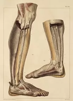
Lower leg and foot fascia, 1831 artwork
Lower leg and foot fascia. Lateral views of the partitions of the fascia of the muscles of the leg and foot. Fascia are fibrous connective tissues that surround muscles and other tissues

Background imageLateral Collection: Foot ligaments and tendons, artwork C016 / 7010

Foot ligaments and tendons, artwork C016 / 7010
Foot ligaments and tendons. Artwork of the bones, ligaments (white, between bones), and tendons (white, muscle-bone) of the foot and the ankle joint from the side

Background imageLateral Collection: Horse anatomy, artwork C016 / 6846

Horse anatomy, artwork C016 / 6846
Horse anatomy. Artwork of the musculature of a horse as seen from the side, including the head, neck, legs, torso and tail

Background imageLateral Collection: Knee muscles and bones, artwork C016 / 7013

Knee muscles and bones, artwork C016 / 7013
Knee muscles and bones. Artwork of an oblique lateral frontal view of the muscles and bones of a human knee. This view is of the outside of the left leg

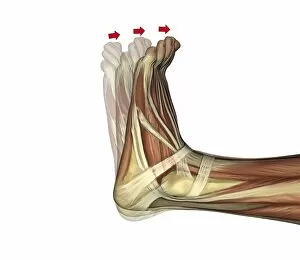
Background imageLateral Collection: Plantarflexion of the foot, artwork C016 / 6800

Plantarflexion of the foot, artwork C016 / 6800
Plantarflexion of the foot. Artwork of the muscles of the foot from the side, with red arrows showing the direction of movement when flexing the foot in the direction of its lower (plantar)

Background imageLateral Collection: Plantarflexion of the foot, artwork C016 / 6799

Plantarflexion of the foot, artwork C016 / 6799
Plantarflexion of the foot. Artwork of the muscles of the foot from the side, with red arrows showing the direction of movement when flexing the foot in the direction of its lower (plantar)

Background imageLateral Collection: Atlanto-occipital joint, artwork C016 / 6557

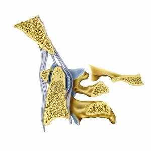
Atlanto-occipital joint, artwork C016 / 6557
Atlanto-occipital joint. Artwork of the bones (yellow) and ligaments (blue) of the atlanto-occipital joint, as seen from the side in a medial sagittal section. The rear of the spine is at right

Background imageLateral Collection: Vertebral joints of the neck, artwork C016 / 6551

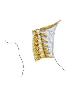
Vertebral joints of the neck, artwork C016 / 6551
Vertebral joints of the neck. Artwork of the bones (yellow) and ligaments (blue) of the cervical (neck) region of the spine (vertebral column)

Background imageLateral Collection: Scoliosis of the spine, X-ray C016 / 6565

Scoliosis of the spine, X-ray C016 / 6565
Scoliosis of the spine. Coloured X-ray of the chest of a 34 year old female patient with scoliosis (sideways curvature) of the spine

Background imageLateral Collection: Dorsiflexion of the foot, artwork C016 / 6797

Dorsiflexion of the foot, artwork C016 / 6797
Dorsiflexion of the foot. Artwork of the muscles of the foot from the side, with red arrows showing the direction of movement when flexing the foot in the direction of its upper (dorsal)

Background imageLateral Collection: Dorsiflexion of the foot, artwork C016 / 6798

Dorsiflexion of the foot, artwork C016 / 6798
Dorsiflexion of the foot. Artwork of the muscles of the foot from the side, with red arrows showing the direction of movement when flexing the foot in the direction of its upper (dorsal)

Background imageLateral Collection: Mouse embryo, prepared specimen C018 / 0318

Mouse embryo, prepared specimen C018 / 0318
Mouse embryo. Macrophotograph of a prepared specimen of a mouse embryo after around 16 days of development. It has been made translucent and its skeleton stained with Alizarin red dye

Background imageLateral Collection: Scoliosis treatment, X-ray C017 / 7156

Scoliosis treatment, X-ray C017 / 7156
Scoliosis treatment. X-ray of the spine of a 46 year old female patient that has been fixed to treat scoliosis (sideways curvature)

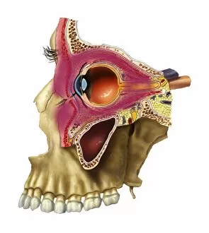
Background imageLateral Collection: Plegmon eye inflammation, artwork

Plegmon eye inflammation, artwork
Plegmon eye inflammation. Artwork of a sagittal section through a human eye and the surrounding structures, in a case of a subcutaneous (below the skin) phlegmon inflammation of an eye cavity

Background imageLateral Collection: Ovarian cyst in abdomen, artwork

Ovarian cyst in abdomen, artwork
Ovarian cyst in abdomen. Artwork of a large ovarian cyst (centre left) in a dissected human abdomen, seen from above. The uterus (centre) and the ovaries are part of the female reproductive system

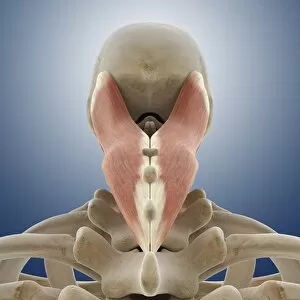
Background imageLateral Collection: Neck muscle, artwork C014 / 5019

Neck muscle, artwork C014 / 5019
Neck muscle. Computer artwork showing the splenius capitis muscle of the neck. This muscle is attached to the upper thoracic (chest) and lower cervical (neck) vertebre and the base of the skull

Background imageLateral Collection: Neck muscle, artwork C014 / 5022

Neck muscle, artwork C014 / 5022
Neck muscle. Computer artwork showing the splenius capitis muscle of the neck. This muscle is attached to the upper thoracic (chest) and lower cervical (neck) vertebre and the base of the skull

Background imageLateral Collection: Neck muscle, artwork C014 / 5018

Neck muscle, artwork C014 / 5018
Neck muscle. Computer artwork showing the splenius capitis muscle of the neck. This muscle is attached to the upper thoracic (chest) and lower cervical (neck) vertebre and the base of the skull

Background imageLateral Collection: Pinned foot bones after surgery, X-ray

Pinned foot bones after surgery, X-ray
Pinned foot bones after surgery. Lateral (side view) X-ray showing pins (white) inserted into the bones of a patients foot

Background imageLateral Collection: Female groin arteries, 1825 artwork

Female groin arteries, 1825 artwork
Female groin arteries. Dissections of a female groin to show areas supplied by the internal iliac artery (red) and its branches

Background imageLateral Collection: Lumbar spinal nerves, 1825 artwork

Lumbar spinal nerves, 1825 artwork
Lumbar spinal nerves. Lateral view of the lower vertebral column, showing nerves (white) of the sympathetic nervous system. The pelvic ilium bone has been removed

Background imageLateral Collection: Lower leg arteries, 1825 artwork

Lower leg arteries, 1825 artwork
Lower leg arteries. Anterior (frontal) view of the arteries (red) and muscles and ligaments of the lower leg and foot. At left are the superficial arteries

Background imageLateral Collection: Lateral trunk muscles, 1831 artwork

Lateral trunk muscles, 1831 artwork
Lateral trunk muscles. Side view (front of body at right) of the third layer of the lateral muscles of the trunk. The muscles include the serratus, the transverse abdominal

Background imageLateral Collection: Femoral and foot arteries, 1825 artwork

Femoral and foot arteries, 1825 artwork
Femoral and foot arteries. Dissections showing the arteries (red) and muscles of the upper thigh and the foot. At centre right, near the bladder (white)

Background imageLateral Collection: Fascia of the thigh, 1831 artwork

Fascia of the thigh, 1831 artwork
Fascia of the thigh. Lateral view (front of body at right) of the external fascia of the muscles of the thigh. Fascia are fibrous connective tissues that surround muscles and other tissues

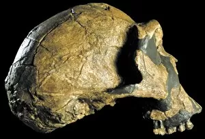
Background imageLateral Collection: Homo ergaster skull (KNM-ER 3733) C015 / 6927

Homo ergaster skull (KNM-ER 3733) C015 / 6927
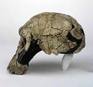
Homo ergaster skull (KNM-ER 3733). This fossil specimen dates from around 1.8 million years ago, and was discovered in 1975 in Koobi Fora, on the eastern shore of Lake Turkana, Kenya

Background imageLateral Collection: Homo ergaster skull (SK-847 and SK-15) C015 / 6928

Homo ergaster skull (SK-847 and SK-15) C015 / 6928
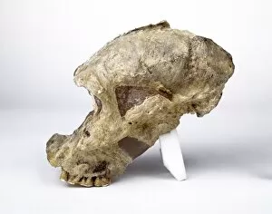
Homo ergaster skull. This skull consists of two fossils: SK-847 (main) and SK-15 (lower jaw). The fossils are those of an extinct species that forms an early part of the human evolutionary tree

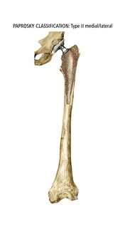
Background imageLateral Collection: Paprosky femur defect, type IIIA med-lat

Paprosky femur defect, type IIIA med-lat
Paprosky femur defect. Cutaway artwork of bone degradation in a type IV lateral femur cortex defect (Paprosky classification system)

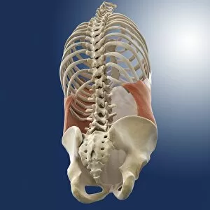
Background imageLateral Collection: Abdominal external oblique muscle C014 / 5062

Abdominal external oblique muscle C014 / 5062
Abdominal external oblique muscle, computer artwork. This thin muscle acts to pull the chest down and compress the abdominal cavity

Background imageLateral Collection: Abdominal external oblique muscle C014 / 5061

Abdominal external oblique muscle C014 / 5061
Abdominal external oblique muscle, computer artwork. This thin muscle acts to pull the chest down and compress the abdominal cavity

Background imageLateral Collection: Fossil sand tiger shark teeth C016 / 5551

Fossil sand tiger shark teeth C016 / 5551
Fossil sand tiger shark teeth (Odontaspis robusta). Anterior teeth (tall, slender) and lateral teeth (triangular) are shown. Found in Early Eocene rocks, Abbey Wood, Kent

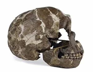
Background imageLateral Collection: Homo neanderthalensis cranium (Tabun 1) C016 / 5201

Homo neanderthalensis cranium (Tabun 1) C016 / 5201
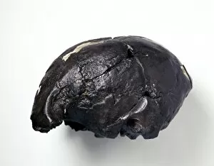
Homo neanderthalensis cranium (Tabun 1). Side view of a cast of a fossil skull of a female Neanderthal (Homo neanderthalensis)

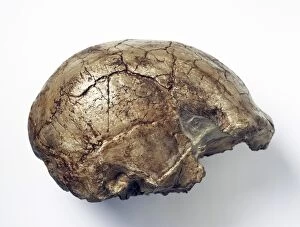
Background imageLateral Collection: Homo sapiens cranium (Omo 1) C016 / 5174

Homo sapiens cranium (Omo 1) C016 / 5174
Homo sapiens cranium (Omo 1). Side view of a reconstruction of the fossil Homo sapiens skull known as Omo 1 and discovered in 1967 by a team led by Richard Leakey

Background imageLateral Collection: Homo sapiens cranium (Omo 2) C016 / 5169

Homo sapiens cranium (Omo 2) C016 / 5169
Homo sapiens cranium (Omo 2). Side view of a cast of a fossil Homo sapiens cranium dating from 130, 000 years ago. Known as Omo 2, it was discovered in 1967 at the Omo River

Background imageLateral Collection: Homo sapiens cranium (LH 18) C016 / 5164

Homo sapiens cranium (LH 18) C016 / 5164
Homo sapiens cranium (LH 18). Side view of a cast of a fossil Homo sapiens cranium known as Laetoli Hominid 18 (LH 18). Dating back 150, 000 years ago

Background imageLateral Collection: Paranthropus robustus cranium (SK46) C016 / 5100

Paranthropus robustus cranium (SK46) C016 / 5100
Paranthropus robustus cranium (SK46). Lateral view of a cast of a fossil of part of the cranium of an adult Paranthropus robustus hominid. This specimen was probably female

Background imageLateral Collection: Taung Child skull (Taung 1) C016 / 5102

Taung Child skull (Taung 1) C016 / 5102
Taung Child skull (Taung 1). Side view of a cast of a fossil skull of an Australopithecus africanus child from Taung, South Africa. A

Background imageLateral Collection: Homo ergaster cranium (KNM-ER 3733) C016 / 5094

Homo ergaster cranium (KNM-ER 3733) C016 / 5094
Homo ergaster skull (KNM-ER 3733). This cast is of a fossil specimen that dates from around 1.8 million years ago, and was discovered in 1975 by Bernard Ngeneo, in Koobi Fora

Background imageLateral Collection: Homo habilis cranium (KNM-ER 1813) C016 / 5091

Homo habilis cranium (KNM-ER 1813) C016 / 5091
Homo habilis cranium (KNM-ER 1813). This cast is of a relatively complete fossil skull named KNM-ER 1813. It was found in Koobi Fora, Kenya in 1973 by Kamoya Kimeu

Background imageLateral Collection: Homo rudolfensis cranium (KNM-ER 1470) C016 / 5088

Homo rudolfensis cranium (KNM-ER 1470) C016 / 5088
Homo rudolfensis cranium (KNM-ER 1470). This fossil specimen dates from around 1.9 million years ago, and was discovered in 1972 in Koobi Fora, on the eastern shore of Lake Turkana, Kenya

Background imageLateral Collection: Homo sapiens skull (Qafzeh 11) C016 / 5081

Homo sapiens skull (Qafzeh 11) C016 / 5081
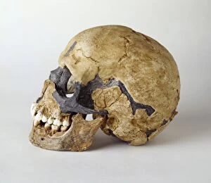
Homo sapiens skull (Qafzeh 11). Cast and reconstruction of the fossil cranium and mandible of the Qafzeh 11 child. Found by Bernard Vandermeersch in 1971, in the Qafzeh cave site in Nazareth, Israel

Background imageLateral Collection: Articular disk of the jaw, artwork C016 / 2889

Articular disk of the jaw, artwork C016 / 2889
Articular disk of the jaw. Artwork of a side view of a human skull with the jaws closed, showing the articular disk (white) and other cartilage of the temporomandibular joint

Background imageLateral Collection: Articular disk of the jaw, artwork C016 / 2890

Articular disk of the jaw, artwork C016 / 2890
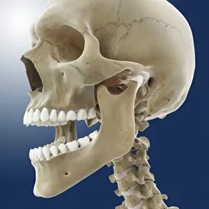
Articular disk of the jaw. Artwork of a side view of a human skull with the jaws open, showing the articular disk (white) and other cartilage of the temporomandibular joint

Background imageLateral Collection: Knee flexion anatomy, artwork C016 / 2880

Knee flexion anatomy, artwork C016 / 2880
Knee flexion anatomy. Artwork of a side (lateral) view of the bones and some of the cartilage and ligaments and tendons of a flexed (left) and extended (right) knee joint


All Professionally Made to Order for Quick Shipping

"Lateral: Exploring the Intricacies of Nature's Design" Delving into the intricate network of brain blood vessels, as captured in a mesmerizing 3D angiogram C007 / 1981. Unveiling the hidden wonders of the human knee joint through an anatomy study that reveals its lateral complexities. Witnessing the majestic Eurasian lynx (Lynx lynx) in all its glory, with its captivating lateral movements and stealthy demeanor. Marveling at the delicate intricacy of brain blood vessels, showcasing nature's remarkable lateral pathways within our most vital organ. Transporting ourselves back to 1956 French Grand Prix, where mechanics examine Trintignants Bugatti Type 251 chassis and its straight 8 Bugatti 251 engine from a lateral perspective. Glimpsing at the striking Red-billed Chough (Pyrrhocorax pyrrhocorax) at Alpine Zoo Innsbruck, Tyrol, Austria, Europe - a testament to nature's lateral adaptations for survival in high altitudes. Admiring an exquisite artwork from 1825 depicting male groin arteries - a fascinating exploration into our body's intricate vascular system from a unique lateral viewpoint. Discovering head and neck muscles through an extraordinary artwork dating back to 1831 - offering insights into their complex interplay and role in our daily movements from various angles including laterally. Examining a tonguestone (sharks tooth) adorned with intriguing lateral denticles that enhance these creatures' feeding efficiency and survival strategies over millions of years. Peering into skull anatomy to unravel mysteries hidden beneath our skin; exploring bone structures and cranial features from both frontal and lateral perspectives for comprehensive understanding. Venturing deep within Earth's core as we explore "Earth's Central Fire, " uncovering geological forces that shape our planet's lateral movements and tectonic shifts.