
Hormones Collection
"Hormones: Unveiling the Intricacies of Athlete Physiology and Artistic Wonders" Delving into the realm of hormones


All Professionally Made to Order for Quick Shipping
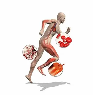
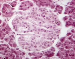
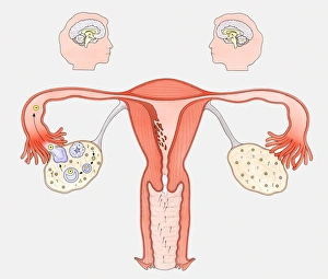
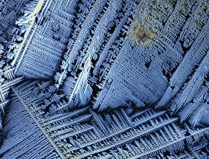
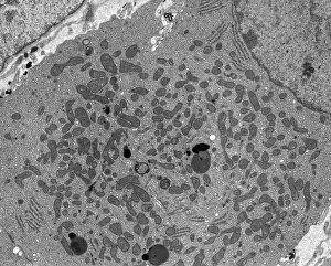
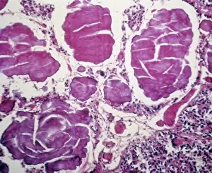
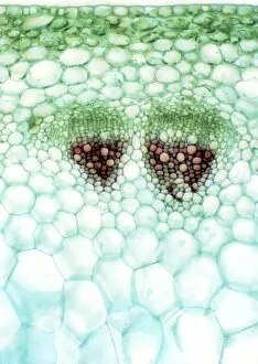
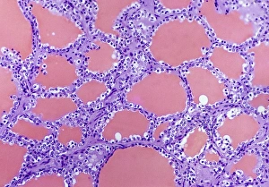
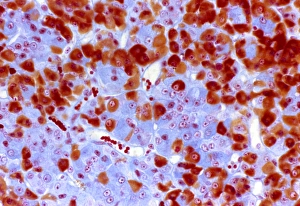
"Hormones: Unveiling the Intricacies of Athlete Physiology and Artistic Wonders" Delving into the realm of hormones, we uncover a fascinating world where athlete physiology intertwines with captivating artwork. From the crystalline structures of testosterone to the mesmerizing light micrograph of Islet of Langerhans, this journey will leave you in awe. Peering through a scanning electron microscope (SEM), we witness the intricate network of capillaries within the thyroid gland, revealing its profound role in hormone regulation. Meanwhile, an enchanting SEM image showcases the delicate blood vessels that nourish this vital gland. Venturing deeper into our brain's limbic system, we unravel how hormones influence our emotions and behaviors. Through stunning artwork depicting Islets of Langerhans cells, we gain insight into their significance in maintaining glucose balance and overall well-being. Intriguingly, history comes alive as an engraving portrays a woman suffering from congenital iodine deficiency syndrome known as cretinism. This poignant reminder highlights the importance of hormonal balance for optimal health. A thought-provoking illustration captures a woman standing confidently while her body faces forward but her head turns to one side. Overlaying this image are skeletal structures and inner organs - a visual representation showcasing how hormones intricately connect every aspect of our being. The complex relationship between female sexual organs and the brain is unveiled through an informative diagram. On one side lies the normal reproductive cycle while on the other side reveals how contraceptive pills alter this natural process – shedding light on modern advancements impacting hormonal equilibrium. Concluding our exploration is another glimpse at Islet of Langerhans through a captivating light micrograph. Its beauty serves as a testament to nature's precision in orchestrating these tiny clusters responsible for regulating blood sugar levels. Hormones transcend mere chemicals; they embody artistry intertwined with athlete physiology—a symphony that orchestrates life's intricate dance.