Fraud Blocker Skip to main content

Histological Collection (page 2)

"Exploring the Intricacies of Histological Wonders: Unveiling the Hidden Beauty within Our Bodies" Step into the fascinating world of histology

Background imageHistological Collection: Heart muscle, confocal light micrograph

Heart muscle, confocal light micrograph
Heart muscle. Confocal light micrograph of a section through cardiac (heart) muscle. Cardiac muscle consists of branching elongated muscle cells

Background imageHistological Collection: Gout crystals

Gout crystals. Polarised light micrograph of uric acid crystals in the synovial fluid (a lubricating liquid in joints) from a patient with gout

Background imageHistological Collection: Plant cell mitosis, light micrograph

Plant cell mitosis, light micrograph
Plant cell mitosis. Light micrograph of root tip cells from an onion (Allium sp.) undergoing cell division (mitosis). From top left to bottom right

Background imageHistological Collection: Thymus gland, light micrograph C015 / 4970

Thymus gland, light micrograph C015 / 4970
Thymus gland. Light micrograph of a section through tissue from the thymus gland, part of the lymphoid system. The dense areas are the thymic cortex, which produces lots of lymphocytes

Background imageHistological Collection: Myeloblast blood cell, light micrograph

Myeloblast blood cell, light micrograph
Myeloblast blood cell. Light micrograph of blood cells, including a myeloblast, a precursor for a type of white blood cell (leucocyte). Myeloblasts differentiate into granulocytes

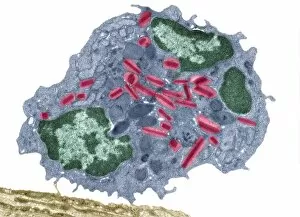
Background imageHistological Collection: Eosinophil white blood cell, TEM

Eosinophil white blood cell, TEM
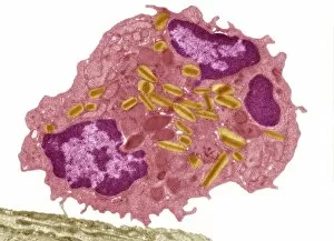
Eosinophil white blood cell. Transmission electron micrograph (TEM) of a section through an eosinophil white blood cell (leukocyte)

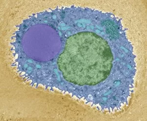
Background imageHistological Collection: Pancreatic acinar cell

Pancreatic acinar cell. Transmission electron micrograph (TEM) of a section through an enzyme-secreting acinar cell in the human pancreas, showing part of the nucleus (round, far left)

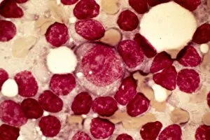
Background imageHistological Collection: Promyelocyte blood cell, light micrograph

Promyelocyte blood cell, light micrograph. This blood cell (centre) is a precursor for a type of white blood cell called a granulocyte, formed by granulopoiesis in the bone marrow

Background imageHistological Collection: Chronic lymphocytic leukaemia, micrograph

Chronic lymphocytic leukaemia, micrograph
Chronic lymphocytic leukaemia. Light micrograph of blood cells sampled from a lymphatic ganglion in a case of chronic lymphocytic leukaemia, also called chronic lymphoid leukaemia (CLL)

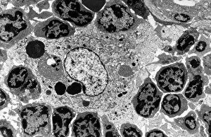
Background imageHistological Collection: Macrophage and lymphocytes, TEM

Macrophage and lymphocytes, TEM
Macrophage and lymphocytes, transmission electron micrograph (TEM). Macrophage surrounded by lymphocytes in a lymph node. This view includes many intercellular contacts

Background imageHistological Collection: Hepatocyte liver cell, TEM

Hepatocyte liver cell, TEM
Hepatocyte liver cell. Transmission electron micrograph (TEM) of a section through an hepatocyte liver cell, showing rough and smooth endoplasmic reticulum (ER, dark lines)

Background imageHistological Collection: Liver tissue, TEM

Liver tissue, TEM
Liver tissue. Transmission electron micrograph (TEM) of a section through the liver, showing part of a radial cord of hepatocyte liver cells (dark) and the vascular sinusoids (white)

Background imageHistological Collection: Cartilage cell, TEM C014 / 1433

Cartilage cell, TEM C014 / 1433
Cartilage cell. Transmission electron micrograph (TEM) of a section through a chondrocyte cell from hyaline cartilage of the trachea (windpipe)

Background imageHistological Collection: Cartilage cell, TEM C014 / 1434

Cartilage cell, TEM C014 / 1434
Cartilage cell. Transmission electron micrograph (TEM) of a section through a chondrocyte cell from hyaline cartilage of the trachea (windpipe)

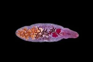
Background imageHistological Collection: Lancet liver fluke C014 / 4846

Lancet liver fluke C014 / 4846
Lancet liver fluke (Dicrocoelium dendriticum). This parasitic trematode (flatworm) tends to live in cattle or other grazing mammals

Background imageHistological Collection: Skeletal muscle, TEM C016 / 5369

Skeletal muscle, TEM C016 / 5369
Skeletal muscle. Transmission electron micrograph (TEM) of a cross section through human skeletal (striated) muscle. Blocks of muscle (lighter grey) are surrounded by connective tissue (black)

Background imageHistological Collection: Earthworm, transverse section

Earthworm, transverse section
Earthworm. Light micrograph of a transverse section through the body of a round segmented earthworm (Lumbricus terrestris) in the intestinal region

Background imageHistological Collection: Bladder epithelium, light micrograph

Bladder epithelium, light micrograph
Bladder epithelium. Light micrograph of a vertical section through the wall of the urinary bladder. The inner surface is at top

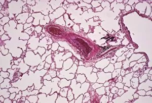
Background imageHistological Collection: Lung alveoli

Lung alveoli. Light micrograph of a section through normal human lung tissue showing alveoli. These are tiny air sacs where most of the diffusion of gases to and from the blood occurs

Background imageHistological Collection: Cardiac muscle, TEM

Cardiac muscle, TEM
Cardiac muscle. Coloured transmission electron micrograph (TEM) of cardiac muscle fibrils (orange)from a healthy heart. Mitochondria (pink) supply the muscle cells with energy

Background imageHistological Collection: Goblet cells

Goblet cells. Coloured transmission electron micrograph (TEM) of a section through goblet cells in the lining of the small intestine, part of the digestive tract. They are full of mucus (yellow)

Background imageHistological Collection: Whale bone tissue, light micrograph

Whale bone tissue, light micrograph

Background imageHistological Collection: Hazel nut, light micrograph

Hazel nut, light micrograph
Hazel nut. Light micrograph of a section through a nut from a common hazel (Corylus avellana) tree, showing the large amount of fat (red) stored within endosperm tissue

Background imageHistological Collection: Desmosome cell junction, artwork

Desmosome cell junction, artwork
Desmosome cell junction. Computer artwork showing the structure of an adhesion junction, or desmosome. Desmosomes form the most common type of junction between epithelial cells

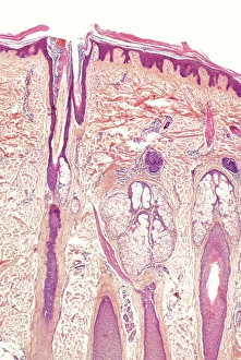
Background imageHistological Collection: Skin and hair follicles, light micrograph

Skin and hair follicles, light micrograph
Skin and hair follicles. Light micrograph of a section through skin, showing hair follicles (dark purple circular structures). Haematoxylin and eosin stain

Background imageHistological Collection: Apple stem, light micrograph

Apple stem, light micrograph
Apple stem. Polarised light micrograph of a section through the stem of a young apple (Malus domestica) tree plant. Magnification: x100 when printed 10 centimetres wide

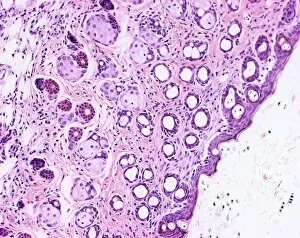
Background imageHistological Collection: Human skin section, light micrograph

Human skin section, light micrograph
Human skin. Light micrograph of a section through healthy human skin. The outer surface of the skin is at top. The uppermost surface is the epidermis

Background imageHistological Collection: Human skin section, light micrograph P710 / 0472

Human skin section, light micrograph P710 / 0472
Human skin. Light micrograph of a section through healthy human skin. The outer surface of the skin is at top. The uppermost surface is the epidermis

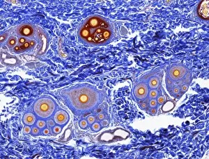
Background imageHistological Collection: Scalp skin showing hair follicles, LM

Scalp skin showing hair follicles, LM
Scalp skin. Light micrograph of a section through skin from the scalp. Numerous hair shafts are seen (purple rings), which are surrounded by connective tissue (dark blue)

Background imageHistological Collection: Eosinophil white blood cell, TEM C014 / 1438

Eosinophil white blood cell, TEM C014 / 1438
Eosinophil white blood cell. Transmission electron micrograph (TEM) of a section through an eosinophil. Eosinophils, like all white blood cells, are part of the bodys immune system

Background imageHistological Collection: Bladder lining, TEM C014 / 1473

Bladder lining, TEM C014 / 1473
Bladder lining. Transmission electron micrograph (TEM) of a section through the epithelial cells that line of a human bladder, showing large flat vesicles (large, blue)

Background imageHistological Collection: Intestinal gland cell, TEM C014 / 1441

Intestinal gland cell, TEM C014 / 1441
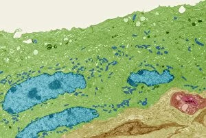
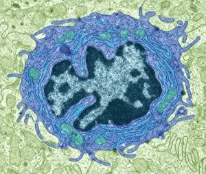
Intestinal gland cell. Transmission electron micrograph (TEM) of a section through an undifferentiated cell from the intestinal tract, showing extensive rough endoplasmic reticulum (ER)

Background imageHistological Collection: Cotton stem, confocal micrograph C014 / 4636

Cotton stem, confocal micrograph C014 / 4636
Cotton stem. Laser scanning confocal micrograph (LSCM) of a section through the stem of a cotton (Gossypium sp.) plant

Background imageHistological Collection: Stem cell-derived retinal cells

Stem cell-derived retinal cells. Fluorescence light micrograph of retinal pigment epithelium (RPE) cells that have been derived from human embryonic stem cells (HESC)

Background imageHistological Collection: Small intestine, light micrograph

Small intestine, light micrograph
Small intestine. Light micrograph of a section through the small intestine. The finger-like projections at left are the villi. These increase the surface area for the absorption of food

Background imageHistological Collection: Stem cell-derived neurons, micrograph

Stem cell-derived neurons, micrograph
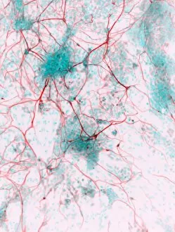
Stem cell-derived neurons. Light micrograph of human nerve cells (neurons) that have been derived from induced pluripotent stem cells (IPS). Tuj1 proteins are cyan, and cell nuclei are red

Background imageHistological Collection: Astrocyte brain cells, light micrograph

Astrocyte brain cells, light micrograph
Astrocyte brain cells. Fluorescence light micrograph of primary astrocyte cells from the brain of a mouse. Astrocytes have numerous branches of connective tissue that provide support

Background imageHistological Collection: Intestinal gland cell, TEM C014 / 1440

Intestinal gland cell, TEM C014 / 1440
Intestinal gland cell. Transmission electron micrograph (TEM) of a section through an undifferentiated cell from the intestinal tract, showing extensive rough endoplasmic reticulum (ER)

Background imageHistological Collection: Cartilage cell, TEM C014 / 1432

Cartilage cell, TEM C014 / 1432
Cartilage cell. Transmission electron micrograph (TEM) of a section through a chondrocyte cell from hyaline cartilage of the trachea (windpipe)

Background imageHistological Collection: Light microscope C014 / 1407

Light microscope C014 / 1407
Light microscope, seen in silhouetted profile. This microscope has a binocular eyepiece (an eyepiece for each eye). The sample is placed on a glass slide on the mount below centre

Background imageHistological Collection: Eosinophil white blood cell, TEM C014 / 1437

Eosinophil white blood cell, TEM C014 / 1437
Eosinophil white blood cell. Transmission electron micrograph (TEM) of a section through an eosinophil. Eosinophils, like all white blood cells, are part of the bodys immune system

Background imageHistological Collection: Brain nerve cells, TEM C014 / 0357

Brain nerve cells, TEM C014 / 0357
Brain nerve cells. Transmission electron micrograph (TEM) of a section through brain tissue from the cerebral cortex, showing numerous neurons (nerve cells) surrounded by axons and dendrites

Background imageHistological Collection: Eosinophil white blood cell, TEM C014 / 1439

Eosinophil white blood cell, TEM C014 / 1439
Eosinophil white blood cell. Transmission electron micrograph (TEM) of a section through an eosinophil. Eosinophils, like all white blood cells, are part of the bodys immune system

Background imageHistological Collection: Golgi apparatus, artwork F006 / 9211

Golgi apparatus, artwork F006 / 9211
Computer artwork of the Golgi apparatus of the human cell. This organelle functions as a central delivery system for the cell

Background imageHistological Collection: Golgi apparatus, artwork F006 / 9199

Golgi apparatus, artwork F006 / 9199
Computer artwork of the Golgi apparatus of the human cell. This organelle functions as a central delivery system for the cell

Background imageHistological Collection: Golgi apparatus, artwork F006 / 9189

Golgi apparatus, artwork F006 / 9189
Computer artwork of the Golgi apparatus of the human cell. This organelle functions as a central delivery system for the cell

Background imageHistological Collection: Stem cell-derived astrocyte brain cells

Stem cell-derived astrocyte brain cells
Stem cell-derived nerve cells. Fluorescence light micrograph of astrocyte brain cells that have been derived from neural (nerve) stem cells from a mouse

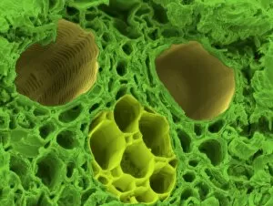
Background imageHistological Collection: Bamboo stem, SEM C015 / 5073

Bamboo stem, SEM C015 / 5073
Bamboo stem. Coloured environmental scanning electron micrograph (ESEM) of a section through the stem of a bamboo plant (family Gramineae), showing a vascular bundle


All Professionally Made to Order for Quick Shipping

"Exploring the Intricacies of Histological Wonders: Unveiling the Hidden Beauty within Our Bodies" Step into the fascinating world of histology, where microscopic wonders come to life. Delve into Santiago Ramon Y Cajal's masterpiece, as his cortical grey matter schema unveils the intricate network that forms our brain's command center. Witness the mesmerizing complexity of a mammalian retina through a histological diagram, revealing how light is transformed into vision. Journey deeper into our cerebellum tissue, as a captivating light micrograph showcases its unique structure and function. Marvel at Ramon Y Cajal's genius once again as he unravels the secrets of synapse nerve junctions through an astonishing TEM image. Venture further into uncharted territory with a glimpse of hippocampus brain tissue, where memories are formed and stored. Observe Purkinje nerve cells in the cerebellum, their elegant arrangement hinting at their crucial role in coordinating movement. Witness nature's precision with kidney tubules in section; these delicate structures filter waste from our bloodstreams tirelessly. Explore human brain microscope slides and be awestruck by its intricate architecture - billions of neurons working harmoniously to shape who we are. Peer closely at rough endoplasmic reticulum through TEM imagery; this cellular powerhouse orchestrates protein synthesis within each living cell. Immerse yourself in glial stem cell culture under a light microscope - witness new life being nurtured for potential regeneration. Finally, marvel at the intricacy of brain tissue blood supply - countless vessels ensuring oxygen and nutrients reach every corner of this remarkable organ. Histological wonders unlock hidden beauty within us all – reminding us that even on a microscopic level, there is awe-inspiring complexity waiting to be discovered.