Fraud Blocker Skip to main content

Eumycota Collection (page 3)

Eumycota, the diverse kingdom of fungi, encompasses a fascinating array of organisms

Background imageEumycota Collection: Cryptococcus cell dividing, TEM

Cryptococcus cell dividing, TEM
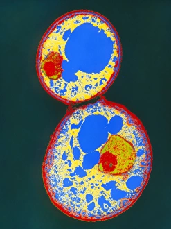
Cryptococcus cell dividing. Coloured transmission electron micrograph (TEM) of the asexual budding (lower right) of a Cyptococcus sp. yeast cell

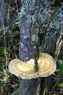
Background imageEumycota Collection: Daedaleopsis confragosa fungus

Daedaleopsis confragosa fungus attached to the trunk of a tree. This bracket fungus grows on dead and living wood of broad leaved trees, particularly alder, birch and willow

Background imageEumycota Collection: Earth star mushroom

Earth star mushroom (Geastrum triplex) growing in woodland soil. It has released (puffed) a cloud of reproductive spores (upper centre)

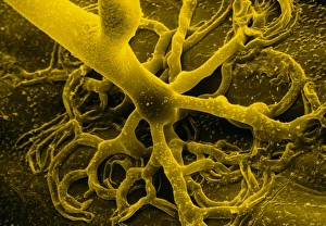
Background imageEumycota Collection: False col SEM of rhizoids

False col SEM of rhizoids
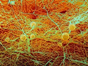
False colour scanning electron micrograph of rhizoids, or " rootlets" of the bread mould, Rhizopus stolonifer, on the surface of bread. Spores circulate freely in the air

Background imageEumycota Collection: False colour SEM of fruiting bodies

False colour SEM of fruiting bodies

Background imageEumycota Collection: Coloured TEM of a yeast cell

Coloured TEM of a yeast cell

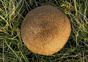
Background imageEumycota Collection: Puffball fungus

Puffball fungus (Lycoperdon foetidum)

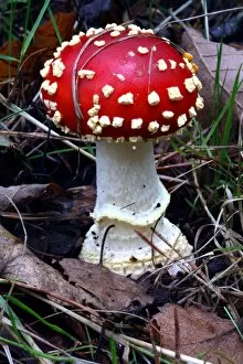
Background imageEumycota Collection: Fly agaric fungus

Fly agaric fungus (Amanita muscaria). The mycelium of the fly agaric fungus shares a symbiotic relationship with the spruce, forming a mycorrhizal sheath around the roots of the tree

Background imageEumycota Collection: Shaggy parasol mushrooms

Shaggy parasol mushrooms
Shaggy parasol mushroom (Macrolepiota rhacodes). This fungus is edible

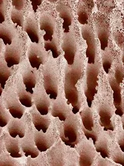
Background imageEumycota Collection: Mushroom surface, SEM

Mushroom surface, SEM
Mushroom surface. Coloured scanning electron micrograph (SEM) of the spore-bearing surface of a hen of the woods mushroom (Grifola frondosa). This surface is found on the underside of the mushroom

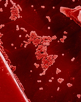
Background imageEumycota Collection: Wine yeast cells, Saccharomyces

Wine yeast cells, Saccharomyces
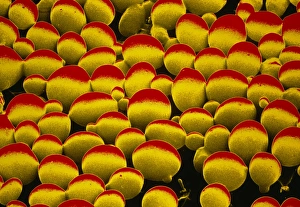
False colour scanning electron micrograph of wine yeast cells Saccharomyces cerevisiae (var. ellipsoideus), (tiny balls) on crystals of potassium hydrogen tartrate precipitated out from red wine &

Background imageEumycota Collection: Cramp balls fungi

Cramp balls fungi (Daldinia concentrica) on a dead ash tree (Fraxinus sp.). This species is inedible

Background imageEumycota Collection: False-col SEM of Penicillium chrysogenum

False-col SEM of Penicillium chrysogenum
False-colour scanning electron micrograph (SEM) of Penicillium chrysogenum, the species of penicillin fungus from which the antibiotic is manufactured

Background imageEumycota Collection: Tooth fungi (Hericium coralloides)

Tooth fungi (Hericium coralloides) on a host tree. The mushroom of this fungi bears its spores on a hymenium (a layer of mother cells)

Background imageEumycota Collection: False-col SEM of yeast cells

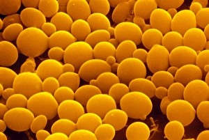
False-col SEM of yeast cells
Yeast. Coloured scanning electron micrograph (SEM) of yeast cells, Saccharomyces cerevisiae, commonly known as Bakers or Brewers yeast, growing on potato dextrose agar

Background imageEumycota Collection: Cauliflower fungus (Sparassis crispa)

Cauliflower fungus (Sparassis crispa) at the base of a tree. This parasite of pine trees causes a brown rot in infected wood

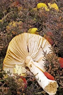
Background imageEumycota Collection: Mushroom gills

Mushroom gills. Macrophotograph of the gills of an unidentified double capped mushroom. Gills are found on the underside of the cap (fruiting body) and hold the fungal spores

Background imageEumycota Collection: Penicillium colony

Penicillium colony. Mat of the fungus Penicillium notatum cultured on a growing medium. Penicillium notatum was the species of fungus which which led Alexander Fleming to discover the antibiotic

Background imageEumycota Collection: Fungus, Penicillium roqueforti

Fungus, Penicillium roqueforti
Macrophotograph (filtered) of Penicillium roqueforti, an economically important penicillin fungus used in the manufacture of blue cheeses

Background imageEumycota Collection: Dryads saddle fungi

Dryads saddle fungi (Polyporus squamosus) on a dead tree stump. This edible fungus is most common in spring and summer. It is parasitic and eventually kills its host

Background imageEumycota Collection: White bread mould, SEM

White bread mould, SEM
White bread mould. Coloured scanning electron micrograph (SEM) of the fruiting bodies of two types of mould growing on white bread. The moulds are Penicillium sp. and Mucor mucedo

Background imageEumycota Collection: Dry rot fungus, SEM

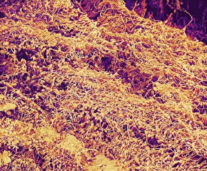
Dry rot fungus, SEM
Dry rot fungus. Coloured scanning electron micrograph (SEM) of dry rot in a piece of domestic plywood. Dry rot is a fungus (Serpula lachrymans) which infects damp timber

Background imageEumycota Collection: Fly agaric (Amanita muscaria)

Fly agaric (Amanita muscaria) mushroom lying among ling heather (Calluna vulgaris). This fungus is poisonous if ingested, however it is often used for its hallucinogenic properties when used in small

Background imageEumycota Collection: Bog beacon fungus (Mitrula paludosa)

Bog beacon fungus (Mitrula paludosa)
Bog beacon fungus (Mitrula paludosa, orange) amongst sphagnum moss in a bog. Photographed in the Cairngorms National Park, Aberdeenshire, Scotland, in June

Background imageEumycota Collection: Death cap fungi

Death cap fungi (Amanita phalloides) at the base of a beech tree (Fagus sp. not seen) The mycelium of the death cap fungus shares a symbiotic relationship with the beech

Background imageEumycota Collection: Sporangium of bread mould, Rhizopus

Sporangium of bread mould, Rhizopus
False colour scanning electron micrograph of a sporangium, or fruiting body, of the common bread mould, Rhizopus stolonifer. Spores circulate freely in the air

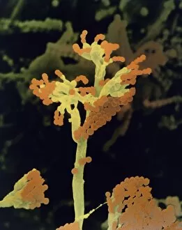
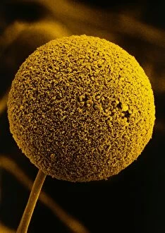
Background imageEumycota Collection: Fungal reproduction, SEM

Fungal reproduction, SEM
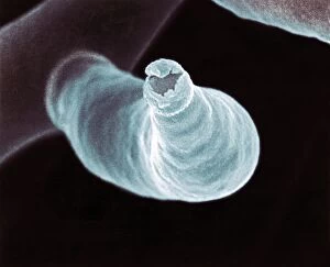
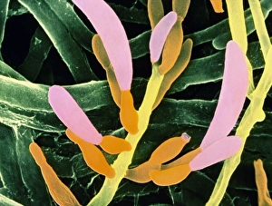
Fungal reproduction. Coloured scanning electron micrograph (SEM) of the tip of a branch of a conidiophore (one type of fungal reproductive structure) with a conidia (spore) emerging from the tip

Background imageEumycota Collection: False-colour SEM of yeast cells

False-colour SEM of yeast cells
Yeast. Coloured scanning electron micrograph of yeast cells, Saccharomyces cerevisiae, commonly known as bakers or Brewers yeast growing on potato dextrose agar

Background imageEumycota Collection: Penicillium fruiting bodies, SEM

Penicillium fruiting bodies, SEM

Background imageEumycota Collection: Shaggy ink cap (Coprinus comatus)

Shaggy ink cap (Coprinus comatus) with black gills. This fungus is deliquescent. It absorbs water molecules from its surrounding environment causing it to become scaly and turn black

Background imageEumycota Collection: Coloured TEM of a budding yeast cell

Coloured TEM of a budding yeast cell
False-colour transmission electron micrograph of a budding yeast cell. Yeast is a kind of single- celled fungus. Budding is a method of asexual reproduction in which a new individual is derived

Background imageEumycota Collection: Bracket polypore fungi

Bracket polypore fungi
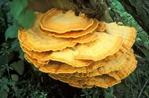
Sulphur polypore or chicken-of-the-woods bracket fungi (Laetiporus sulphureus) on a live willow tree (Salix sp.). This edible fungus is named after the sulphur colour of its hymenium

Background imageEumycota Collection: Gem-studded puffball

Gem-studded puffball fungi (Lycoperdon perlatum). These fungi are white in colour when young and turn brown once mature. They are only edible when they are young

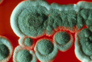
Background imageEumycota Collection: Colonies of Penicillium chrysogenum fungus

Colonies of Penicillium chrysogenum fungus
Macrophotograph of colonies of the fungus Penicillium chrysogenum, which is used in the commercial production of antibiotics

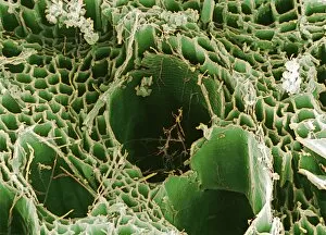
Background imageEumycota Collection: Rotten wood, SEM

Rotten wood, SEM
Rotten wood. Coloured scanning electron micrograph (SEM) of a piece of domestic plywood infected with dry rot fungus. The structure of the wood is seen here

Background imageEumycota Collection: Oyster mushrooms

Oyster mushrooms (Pleurotus ostreatus) growing on a tree trunk. These fruiting bodies (reproductive structures) are produced year-round by the fungus in large clusters

Background imageEumycota Collection: Scarlet elf cup fungi

Scarlet elf cup fungi (Sarcoscypha austriaca) on dead wood. This edible fungus grows in spring on dead rotting branches and in boggy alder wood areas

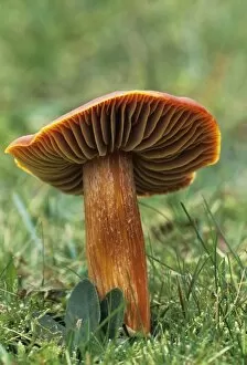
Background imageEumycota Collection: Crimson wax cap mushroom

Crimson wax cap mushroom (Hygrocybe punicea). Photographed in the New Forest, UK

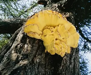
Background imageEumycota Collection: Sulphur polypore fungus on tree

Sulphur polypore fungus on tree
The sulphur polypore, or chicken-of-the-woods fungus, Laetiporus sulphureus, growing on the trunk of a larch tree. An edible fungus when young & fresh, it is considered a delicacy in Germany

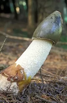
Background imageEumycota Collection: Stinkhorn fungus

Stinkhorn fungus (Phallus impudicus) recently emerged from the egg stage. The tip of the fungus exudes a glutinous gleba (spore mass)

Background imageEumycota Collection: False-colour SEM of bread mould

False-colour SEM of bread mould
False-colour scanning electron micrograph of a bread mould, Mucor mucedo, growing on the surface of a slice of bread. Spores circulate freely in the air

Background imageEumycota Collection: Waxcap fungi

Waxcap fungi (Hygrocybe laeta) in grassland which has not been subjected to artificial fertiliser. Photographed in Abergavenny, Wales, in November

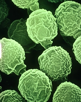
Background imageEumycota Collection: False-colour SEM of the fungus Fusarium oxysporum

False-colour SEM of the fungus Fusarium oxysporum
False-colour scanning electron micrograph of the pathogenic fungus Fusarium oxysporum, which causes wilt disease in tomato and carnation plants

Background imageEumycota Collection: Bread mould, Rhizopus

Bread mould, Rhizopus
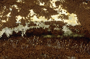
Macrophotograph of a thirteen day old growth of a bread mould (fungus) Rhizopus sp. The slices of bread are overgrown with a network of vegetative hyphae (transparent & threadlike)

Background imageEumycota Collection: Wolfs udder fungus (Lycogala epidendrum)

Wolfs udder fungus (Lycogala epidendrum). Also called wolfs milk, this is not actually a fungus but a slime mould. Photographed growing amongst moss on a Scots pine stump, Cairngorms National Park

Background imageEumycota Collection: Fairy ring fungus

Fairy ring fungus (Marasmius oreades) in a grassy field. This fungus produces a characteristic ring of brown, rounded mushrooms after heavy rainfall in summer and autumn

Background imageEumycota Collection: Meadow waxcap fungi

Meadow waxcap fungi (Hygrocybe pratensis) in grass. This fungus is edible

Background imageEumycota Collection: Stinky squid fungus

Stinky squid fungus


All Professionally Made to Order for Quick Shipping

Eumycota, the diverse kingdom of fungi, encompasses a fascinating array of organisms. From the microscopic world of budding yeast cells and dividing yeast cells seen through scanning electron microscopy (SEM), to the enchanting fly agaric mushrooms with their distinctive red caps and white spots, this caption explores some captivating members of Eumycota. In our first glimpse into this kingdom, we encounter the SEM image of penicillin fungus - a remarkable organism that has revolutionized medicine by producing antibiotics. Moving on to Aspergillus nidulans fungus in culture, its intricate structures captivate us as it thrives in laboratory conditions. Nature's artistry is showcased through the Cep mushroom or Boletus edulis - an edible delicacy prized for its rich flavor and meaty texture. The SEM image of mushroom gills reveals their intricate network responsible for spore production and dispersal. Fly agaric fungi transport us into fairy tales with their vibrant colors and whimsical appearance. These iconic mushrooms have long been associated with folklore and mythical creatures. Returning to the microscopic realm, we witness dividing yeast cells under SEM once again - a testament to their incredible ability to reproduce rapidly. Penicillium roqueforti fungus takes center stage next; known for its role in aging blue cheeses like Roquefort, it adds distinct flavors as it grows within these culinary delights. Fruiting bodies of Rhizopus oligosporus catch our attention next – these structures are essential for reproduction in this species while Cryptococcus neoformans fungi reveal themselves under light microscopy (LM). This pathogenic fungus can cause severe infections particularly in individuals with compromised immune systems. Eumycota truly showcases nature's diversity at both macroscopic and microscopic levels. Whether they provide sustenance or pose health risks, these organisms remind us how intricately interconnected our world is with fungi playing vital roles across various ecosystems.