
Deformity Collection (#6)
"Embracing the Uniqueness: Exploring the Complexity of Deformity" In a world where beauty standards often prevail


All Professionally Made to Order for Quick Shipping
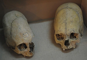
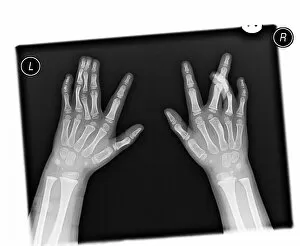
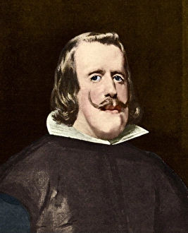
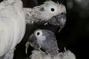
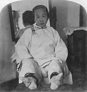
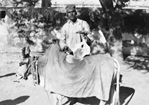
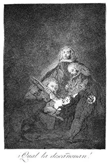
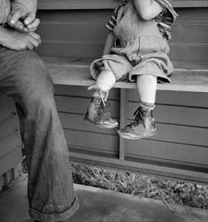
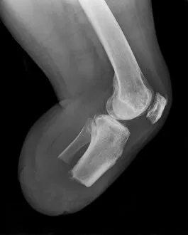
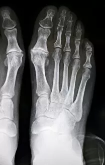
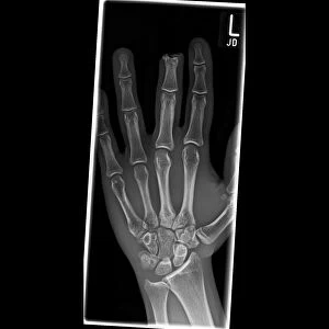
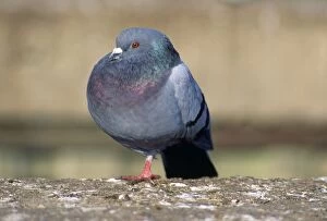
"Embracing the Uniqueness: Exploring the Complexity of Deformity" In a world where beauty standards often prevail, deformities challenge our perceptions and push us to question what it truly means to be normal. From bunions causing discomfort with every step to X-rays revealing intricate skeletal abnormalities, deformity manifests itself in various forms. Take John Baggridge, affectionately known as Jack Double-head. His physical appearance may have been unconventional, but his spirit shone through his unique features. Similarly, a coloured MRI brain scan unveils the complexities of Sturge-Weber syndrome, reminding us that beneath the surface lies an intricate tapestry of human diversity. Nature too holds its share of anomalies; like the Greater bulldog bat captured in Surama, Guyana. Its portrait showcases how even within species, individual differences can arise – a testament to nature's boundless creativity. Moving beyond creatures and humans alike, we encounter historical artifacts such as the Stele of Roma dedicated to Goddess Astarte. This ancient relic reminds us that throughout history people have sought solace and protection from higher powers despite their physical imperfections. Artistic expressions also shed light on society's perception of deformity. The engraving depicting the deformed woman of Prague in 1596 serves as a haunting reminder that prejudice has long plagued humanity. Meanwhile, Tristan and Isalde find love amidst adversity in an 1837 engraving by Baumgarten - showcasing how beauty transcends physical appearances. Throughout literature and folklore too we find tales exploring deformity's impact on individuals' lives - Edgar Allan Poe's "Hop-Frog" delves into themes of revenge fueled by societal ridicule while "The Rhinegold and Valkyrie" illustrates mime writhing under lashes received due to his unusual appearance. Even explorations across continents reveal encounters with difference.