Fraud Blocker Skip to main content

Clinical Collection (page 2)

"Unveiling the Clinical World: From Soviet Innovations to Modern Medical Marvels" Step into the world expertise

Background imageClinical Collection: Silver vaccinating lancet, circa 1870 C017 / 0708

Silver vaccinating lancet, circa 1870 C017 / 0708
Silver plated vaccinating lancet by J. H. Montague, London. J. H. Montague was a British manufacturer of medical instruments, his name can be seen engraved on the handle

Background imageClinical Collection: Chemical reaction F006 / 7185

Chemical reaction F006 / 7185
Chemical reaction. Red liquid boiling inside a flask

Background imageClinical Collection: Pocket monaural stethoscope, circa 1870 C017 / 6957

Pocket monaural stethoscope, circa 1870 C017 / 6957
Three-piece pocket monaural stethoscope, made from ivorine and nickel. French physician, Rene Theophile Hyacinthe Laennec originally invented the monaural stethoscope in 1816

Background imageClinical Collection: Monaural stethoscope, circa 1870 C017 / 6975

Monaural stethoscope, circa 1870 C017 / 6975
Monaural stethoscope dating from 1870. The stethoscope is made of gutta percha with a rubber ring at the earpiece end which also functions as a percussor

Background imageClinical Collection: Monaural stethoscope, circa 1870 C017 / 6937

Monaural stethoscope, circa 1870 C017 / 6937
Billings type monaural stethoscope, made out of fruitwood, dating from around 1870, England. French physician, Rene Theophile Hyacinthe Laennec invented the monaural stethoscope in 1816

Background imageClinical Collection: Monaural stethoscope, circa 1820 C017 / 6930

Monaural stethoscope, circa 1820 C017 / 6930
English monaural stethoscope, made out of fruitwood, dating from the year 1820. French physician, Rene Theophile Hyacinthe Laennec invented the monaural stethoscope in 1816

Background imageClinical Collection: Phonendoscope, circa 1900 C017 / 6968

Phonendoscope, circa 1900 C017 / 6968
Phonendoscope dating from around 1900, England. This device was used in the early 20th century for listening to internal sounds of the body (auscultation)

Background imageClinical Collection: Drainage trocar with cannulae, circa 1860 C017 / 6950

Drainage trocar with cannulae, circa 1860 C017 / 6950
Dr. Southeys drainage trocar with 3 cannulae arranged on an orange background. This set dates from 1860 and is made by Arnold and Sons, England. The instruments are made from ivory and silver

Background imageClinical Collection: Inflamed kidney

Inflamed kidney
Inflamed and swollen kidney, gross specimen. This is a case of chronic nephritis, an inflammation of the kidney. Nephritis is often caused by a bacterial infection

Background imageClinical Collection: Polycystic kidney

Polycystic kidney. Gross specimen of a polycystic kidney, where most of the tissue of the kidneys has been replaced by cysts

Background imageClinical Collection: Bladder adhesions

Bladder adhesions. Gross specimen of a urinary bladder with adhesions (string-like) across its inner surface. Adhesions are growths of fibrous (scar)
Background imageClinical Collection: Phonendoscope, circa 1900 C017 / 6962

Phonendoscope, circa 1900 C017 / 6962
Phonendoscope in its case dating from around 1900, England. This device was used in the early 20th century for listening to internal sounds of the body (auscultation)
Background imageClinical Collection: Phonendoscope, circa 1900 C017 / 6964

Phonendoscope, circa 1900 C017 / 6964
Phonendoscope dating from around 1900, England. This device was used in the early 20th century for listening to internal sounds of the body (auscultation)

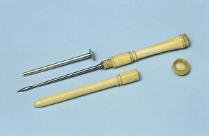
Background imageClinical Collection: Trocar and cannula, circa 1860 C017 / 6960

Trocar and cannula, circa 1860 C017 / 6960
Trocar and cannula dating from 1860, England. The instruments are made from ivory, steel and silver. They were generally used to drain fluid-filled swellings

Background imageClinical Collection: Monaural stethoscope, circa 1860 C017 / 6974

Monaural stethoscope, circa 1860 C017 / 6974
Monaural stethoscope, designed by French doctor, Pierre Piorry, dating from 1860. The chest piece (bell shaped end) and stem are made from fruitwood and the earpiece is made from ebony

Background imageClinical Collection: Skin thermometer, circa 1890 C017 / 0752

Skin thermometer, circa 1890 C017 / 0752
Immisch patent silver skin thermometer in its case. German born Moritz Immisch was a well known clinical instrument maker and obtained the patent for this small, watch-shaped thermometer in 1881

Background imageClinical Collection: Three monaural stethoscopes, circa 1870 C017 / 6956

Three monaural stethoscopes, circa 1870 C017 / 6956
Three two-piece monaural stethoscopes made from ivorine and nickel (left), ebony (centre) and gutta percha and rubber (right)

Background imageClinical Collection: Piorry monaural stethoscope, circa 1850 C017 / 6970

Piorry monaural stethoscope, circa 1850 C017 / 6970
Monaural stethoscope, designed by French doctor, Pierre Piorry, dating from 1850. The chest piece (bell shaped end) and ear piece (thinner end)

Background imageClinical Collection: Etui with syringe, 19th century C017 / 0732

Etui with syringe, 19th century C017 / 0732
Silver plated etui (ornamental case) containing hypodermic syringe and bottle. This etui, engraved with a floral pattern is English and dates from around 1880

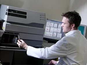
Background imageClinical Collection: Scientist loading a centrifuge

Scientist loading a centrifuge
MODEL RELEASED. Scientist loading a sample into a centrifuge

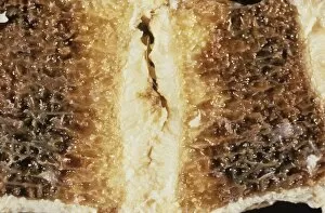
Background imageClinical Collection: Backbone fat deposition C015 / 6053

Backbone fat deposition C015 / 6053
Backbone fat deposition. Sectioned gross clinical specimen of a vertebra (spinal bone) that displays adiposis (fat deposition, yellow) in the medullary (inner) region

Background imageClinical Collection: Spleen cancer, gross specimen C015 / 6421

Spleen cancer, gross specimen C015 / 6421
Spleen cancer. Gross specimen of a spleen showing a large lymphosarcoma, or non-Hodgkins lymphoma, a cancer of lymphoid tissue. The lymphatic system is a major part of the immune system

Background imageClinical Collection: Two praying mantises

Two praying mantises
Praying mantises (Choeradodis rhomboidea). These specimens, from French Guiana, South America, have been preserved so that their haemolymph (body fluid) can be extracted

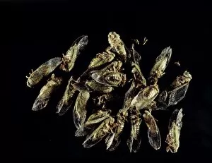
Background imageClinical Collection: Cicada insects

Cicada insects (Carineta aratayensis). These specimens have been preserved so that their haemolymph (body fluid) can be extracted

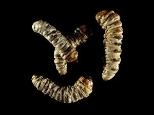
Background imageClinical Collection: Castniid moth larvae

Castniid moth larvae. These specimens, from French Guiana in South America, have been preserved so that their haemplymph (body fluid) can be extracted

Background imageClinical Collection: Chrysomelid beetles

Chrysomelid beetles (Platyphora aulica). These specimens have been preserved so that their haemolymph (body fluid) can be preserved

Background imageClinical Collection: Blood group A

Blood group A. Computer artwork of red blood cells (erythrocytes) in the shape of the letter A. This represents blood group A

Background imageClinical Collection: Blood analysis

Blood analysis. Doctor using a light microscope to analyse a sample of blood. Blood disorders that can be diagnosed this way include malaria. Photographed the Gulu region of Uganda

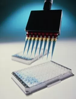
Background imageClinical Collection: Researcher using multichannel pipette, ELISA test

Researcher using multichannel pipette, ELISA test

Background imageClinical Collection: Blood groups

Blood groups. Computer artwork of red blood cells (erythrocytes) and the letters A, B, AB and O. This represents the four different blood groups

Background imageClinical Collection: Blood group AB

Blood group AB. Computer artwork of red blood cells (erythrocytes) in the shape of the letters A and B. This represents blood group AB

Background imageClinical Collection: ELISA blood test for antibodies to disease

ELISA blood test for antibodies to disease
Blood test. Multi-pipette puts blood samples into a multi-well sample tray during an Enzyme-Linked ImmunoSorbent Assay (ELISA)

Background imageClinical Collection: Blood test results

Blood test results. Medical technician examining the printed results from a blood test. The blood sample has been analysed by this automated machine

Background imageClinical Collection: Blood group O

Blood group O. Computer artwork of red blood cells (erythrocytes) in the shape of the letter O. This represents blood group O

Background imageClinical Collection: Blood sample testing

Blood sample testing. Medical technician preparing blood samples for a chemiluminescence immunity test. This test uses fluorescent antibodies to bind to antigens in the blood

Background imageClinical Collection: Blood group B

Blood group B. Computer artwork of red blood cells (erythrocytes) in the shape of the letter B. This represents blood group B

Background imageClinical Collection: Stem cell blood analysis

Stem cell blood analysis
MODEL RELEASED. Stem cell blood analysis. Technician using a flow cytometer to examine a sample of blood stem cells. The cells have been tagged with a fluorescent antibody to a surface protein (CD 34)

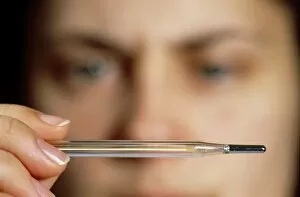
Background imageClinical Collection: Reading of a clinical thermometer

Reading of a clinical thermometer

Background imageClinical Collection: Haemolymph samples

Haemolymph samples in Eppendorf tubes. Haemolymph (body fluid) is obtained from the body cavity (coelom) of insects that have been exposed to bacteria and fungi to trigger an immune response

Background imageClinical Collection: Medical records

Medical records. Nurse carrying a pile of medical records. This could represent the burden of paperwork in hospitals

Background imageClinical Collection: Blood test

Blood test. Drop of blood on a watchglass. Beneath the watchglass is a blood test result sheet

Background imageClinical Collection: Blood analysis, conceptual artwork

Blood analysis, conceptual artwork
Blood testing, conceptual artwork. A syringe is used to take a sample of blood for testing. At upper left are the levels of some blood cells in the tested blood

Background imageClinical Collection: Blood testing

Blood testing
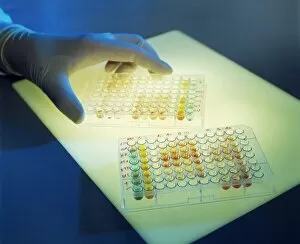
Blood analysis. Gloved hand with multi-well plates containing blood for analysis. Blood can be analysed to check for the presence or absence of antigens, allergens, drugs or pathogenic organisms

Background imageClinical Collection: AIDS blood testing

AIDS blood testing
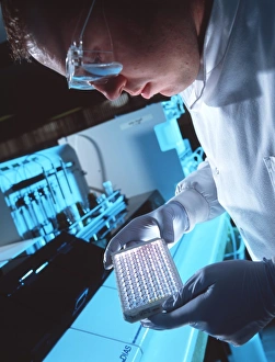
MODEL RELEASED. AIDS blood test. Technician holding a multi-well sample tray as he tests blood for HIV. HIV (human immunodeficiency virus) causes AIDS (acquired immune deficiency syndrome)

Background imageClinical Collection: Ruptured heart

Ruptured heart. Gross specimen of a heart that has ruptured in the parietal region (dark blood clots, lower centre, and red region, centre)

Background imageClinical Collection: Armand Trousseau, French physician

Armand Trousseau, French physician

Background imageClinical Collection: Blood research

Blood research. Pool of blood in a petri dish (out of focus)

Background imageClinical Collection: Pipetting blood

Pipetting blood into a sample pot of clear liquid


All Professionally Made to Order for Quick Shipping

"Unveiling the Clinical World: From Soviet Innovations to Modern Medical Marvels" Step into the world expertise, where hospital nurses don warning jackets as guardians of patient well-being. Inspired by Konstantin Buteyko, the visionary Soviet doctor who revolutionized respiratory therapy, they strive for excellence in every breath. In this realm of healing, Valdecoxib emerges as a potent anti-inflammatory drug, offering relief and hope to those battling pain. Joseph and Louisa Rhine's pioneering research on parapsychology adds an intriguing dimension to clinical exploration, delving into the mysteries of the human mind. Witness a doctor's skilled hands performing a thoracic and abdominal recognition on a patient - a delicate dance between science and compassion. Stephenson Pamela Young's smiling blonde presence radiates warmth amidst these clinical surroundings. Enter an operating theater that hums with focused energy; JLP01_10_54906 captures moments when lives are transformed through surgical precision. A nurse washing her hands in 1928 reminds us of timeless hygiene practices that safeguard against infections. Travel back further in time to witness "A Clinical Lesson with Doctor Charcot at the Salpetriere" - an oil painting from 1887 immortalizing medical education steeped in observation and enlightenment. Men and women diligently working at R. Martens & Co. , Inc. 's export office symbolize meticulous attention to detail essential for effective healthcare systems. Marvel at Florence's Pellizzari Institute room from 1905 - its walls bear witness to physioradiotherapeutic therapy advancements that pushed boundaries towards holistic healing approaches. Finally, explore scientific laboratories adorned with chemical signs from Encyclopedia Britannica – symbols representing knowledge-seeking minds dedicated to unraveling medical complexities. These glimpses into the vast tapestry endeavors remind us that behind every innovation lies countless hours of dedication by passionate individuals striving for better health outcomes worldwide.