Fraud Blocker Skip to main content

Central Nervous System Collection (page 2)

The central nervous system, the intricate network that governs our every thought and movement, is a marvel of complexity

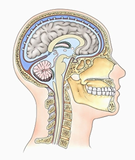
Background imageCentral Nervous System Collection: Lateral cross section through the skull showing the brain with intermediate layers (meninges)

Lateral cross section through the skull showing the brain with intermediate layers (meninges) highlighted

Background imageCentral Nervous System Collection: Medical chart showing the signs and symptoms of sciatica

Medical chart showing the signs and symptoms of sciatica

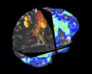
Background imageCentral Nervous System Collection: Brain tumour, fMRI and tractography C017 / 7102

Brain tumour, fMRI and tractography C017 / 7102
Brain tumour, fMRI and tractography. Combined functional magnetic resonance imaging (fMRI, blue and green) and tractography (yellow and red) imaging of a brain with a tumour (upper left)

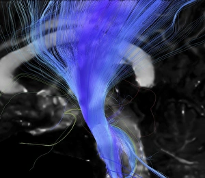
Background imageCentral Nervous System Collection: Corticospinal tract, DTI MRI scan C017 / 7046

Corticospinal tract, DTI MRI scan C017 / 7046
Corticospinal tract. Sagittal 3D diffusion tensor imaging (DTI) magnetic resonance imaging (MRI) scan of nerve pathways (blue) in the brains corticospinal tract

Background imageCentral Nervous System Collection: Normal human brain, MRI scan C016 / 8843

Normal human brain, MRI scan C016 / 8843
Brain. Coloured magnetic resonance imaging (MRI) scan of a sagittal section through a healthy human brain. The highly folded area (brown) is the cerebrum, which is responsible for conscious thought

Background imageCentral Nervous System Collection: Brain blood vessels, artwork

Brain blood vessels, artwork
Brain blood vessels. Computer artwork showing the blood vessels that supply the human brain. The front of the brain is at left

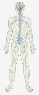
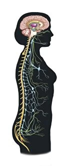
Background imageCentral Nervous System Collection: Illustration of human Central Nervous System showing brain, spinal cord, veins and arteries

Illustration of human Central Nervous System showing brain, spinal cord, veins and arteries

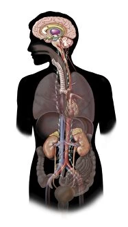
Background imageCentral Nervous System Collection: Human body showing autonomic nervous system and limbic system

Human body showing autonomic nervous system and limbic system
Side view of human body showing autonomic nervous system and limbic system within the brain. Green are parasympathetic nerves. Blue are sympathetic nerves

Background imageCentral Nervous System Collection: Brain surface anatomy, with labels

Brain surface anatomy, with labels

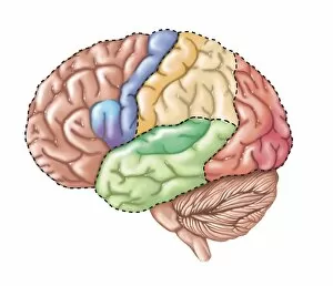
Background imageCentral Nervous System Collection: Side view of the human brain showing the functional lobes

Side view of the human brain showing the functional lobes

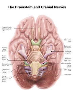
Background imageCentral Nervous System Collection: Anatomy of human brain stem and cranial nerves

Anatomy of human brain stem and cranial nerves

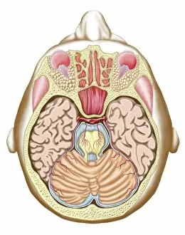
Background imageCentral Nervous System Collection: Transverse section of the midbrain

Transverse section of the midbrain

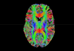
Background imageCentral Nervous System Collection: Brain fibres, tract density imaging C017 / 7039

Brain fibres, tract density imaging C017 / 7039
Brain fibres, tract density imaging. Axial scan of the brain showing the density of nerve pathways (tracts) in the brain. The image was obtained using 3D diffusion tensor imaging (DTI)

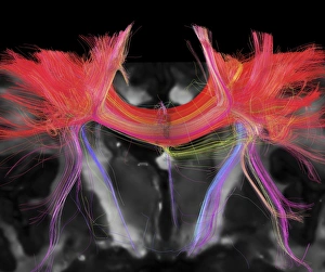
Background imageCentral Nervous System Collection: Corpus callosum, DTI MRI scan C017 / 7043

Corpus callosum, DTI MRI scan C017 / 7043
Corpus callosum. Coronal 3D diffusion tensor imaging (DTI) magnetic resonance imaging (MRI) scan of nerve pathways (red) in the brains corpus callosum

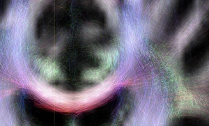
Background imageCentral Nervous System Collection: Brains white matter, DTI MRI scan

Brains white matter, DTI MRI scan
Brains white matter. Close-up of an area of the brain imaged using tract density imaging and 3D diffusion tensor imaging (DTI), a magnetic resonance imaging (MRI) technique

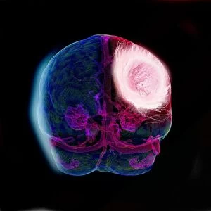
Background imageCentral Nervous System Collection: Brain tumour, DTI modelling C017 / 7060

Brain tumour, DTI modelling C017 / 7060
Brain tumour. Computer artwork of nerve pathways (coloured) in a brain with a tumour (white/red, upper right). The brain is seen from the front. Brain tumours can be benign or malignant (cancerous)

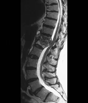
Background imageCentral Nervous System Collection: Tuberculosis of the spine, MRI scan

Tuberculosis of the spine, MRI scan
Tuberculosis of the spine. Magnetic resonance imaging (MRI) scan of a sagittal section through the spine of a 74-year-old patient with tuberculosis (TB) of the spine, or Potts disease

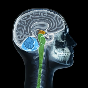
Background imageCentral Nervous System Collection: Human head, X-ray artwork

Human head, X-ray artwork
Human head. Computer artwork of an X-ray through a human head, showing the brain (grey, top), cerebellum (blue), and brainstem and spinal cord (green)

Background imageCentral Nervous System Collection: Brain fibres, DTI MRI scan C017 / 7036

Brain fibres, DTI MRI scan C017 / 7036
Brain fibres. 3D diffusion tensor imaging (DTI) magnetic resonance imaging (MRI) scan of a selection of nerve pathways (green/yellow) in the brain

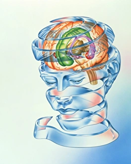
Background imageCentral Nervous System Collection: Brain limbic system

Brain limbic system
Limbic system. Artwork of the limbic system of the brain, a network involved in the expression of instinct and mood. The hippocampus (large purple oval, upper right) is involved in memory

Background imageCentral Nervous System Collection: Brain meninges

Brain meninges, historical anatomical artwork. This cranial (top) view of the brain (with front at the top) and its surrounding protective meninges, which have been partially dissected away

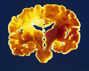
Background imageCentral Nervous System Collection: Coronal slice through a healthy human brain

Coronal slice through a healthy human brain
Human brain. Vertical slice through a healthy brain, sectioned coronally from left to right. This cut in the middle of the brain exposes major anatomical regions

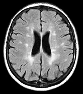
Background imageCentral Nervous System Collection: Multiple sclerosis, MRI scan

Multiple sclerosis, MRI scan

Background imageCentral Nervous System Collection: Brain tumour, 3D-MRI scan

Brain tumour, 3D-MRI scan
Brain tumour. Coloured three-dimensional model of a male head, made up from numerous magnetic resonance imaging (MRI) scans

Background imageCentral Nervous System Collection: Illustration of bundle of nerves in human spine

Illustration of bundle of nerves in human spine

Background imageCentral Nervous System Collection: Illustration of Oligodendrocyte glia cell or neuroglia

Illustration of Oligodendrocyte glia cell or neuroglia

Background imageCentral Nervous System Collection: Conceptual image of synapse of neuron inside brain

Conceptual image of synapse of neuron inside brain

Background imageCentral Nervous System Collection: Microscopic view of multiple nerve cells, known as neurons

Microscopic view of multiple nerve cells, known as neurons
Microscopic view of multiple nerve cells, which are also called neurons. These are responsible for passing information around the central nervous system within the human body

Background imageCentral Nervous System Collection: Illustration of muscle contraction

Illustration of muscle contraction

Background imageCentral Nervous System Collection: Microscopic view of a unipolar neuron

Microscopic view of a unipolar neuron. A unipolar neuron is a type of neuron in which only one protoplasmic process (neurite) extends from the cell body

Background imageCentral Nervous System Collection: Brain surface anatomy

Brain surface anatomy

Background imageCentral Nervous System Collection: Detail of a nerve bundle

Detail of a nerve bundle

Background imageCentral Nervous System Collection: Sciatica caused from herniated disc

Sciatica caused from herniated disc

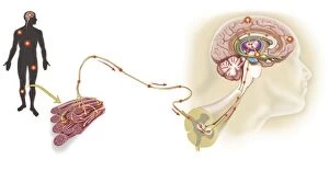
Background imageCentral Nervous System Collection: Pathway of a pain message via sensory nerve in injured muscle

Pathway of a pain message via sensory nerve in injured muscle, to pain gate in spinal cord to limbic system, frontal cortex and sensory cortex in the human brain

Background imageCentral Nervous System Collection: Anatomy of human nervous system and lymphatic system

Anatomy of human nervous system and lymphatic system, front view

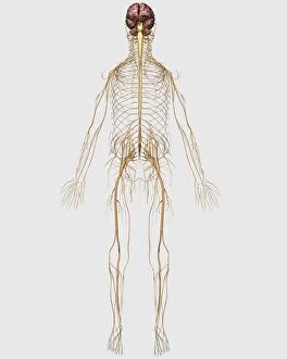
Background imageCentral Nervous System Collection: Medical illustration of peripheral nervous system with brain

Medical illustration of peripheral nervous system with brain

Background imageCentral Nervous System Collection: Anatomy of pituitary gland

Anatomy of pituitary gland

Background imageCentral Nervous System Collection: Representation of how our senses affect our thoughts

Representation of how our senses affect our thoughts

Background imageCentral Nervous System Collection: Conceptual image of synaptic vesicles

Conceptual image of synaptic vesicles

Background imageCentral Nervous System Collection: Conceptual image of human brain with neurons

Conceptual image of human brain with neurons in background

Background imageCentral Nervous System Collection: Representation of how our thoughts affect our emotions

Representation of how our thoughts affect our emotions

Background imageCentral Nervous System Collection: A nerve synapse showing the release of neurotransmitters

A nerve synapse showing the release of neurotransmitters
Detail of a nerve synapse showing the release of neurotransmitters

Background imageCentral Nervous System Collection: Microscopic view of nerve fibers

Microscopic view of nerve fibers. A nerve fiber is a threadlike extension of a nerve cell in the nervous system

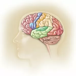
Background imageCentral Nervous System Collection: Side view of the human head showing the functional lobes of the brain

Side view of the human head showing the functional lobes of the brain
Side view of the human head showing the functional lobes of the human brain

Background imageCentral Nervous System Collection: The sympathetic nervous system and the organs of fight-or-flight response

The sympathetic nervous system and the organs of fight-or-flight response

Background imageCentral Nervous System Collection: View of limbic system as seen from directly above the head

View of limbic system as seen from directly above the head

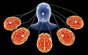
Background imageCentral Nervous System Collection: Conceptual image of multi-brain processing

Conceptual image of multi-brain processing

Background imageCentral Nervous System Collection: Anatomy structure of neurons

Anatomy structure of neurons


All Professionally Made to Order for Quick Shipping

The central nervous system, the intricate network that governs our every thought and movement, is a marvel of complexity. From the delicate cerebellum tissue to the detailed light micrograph capturing its essence, we are reminded of its importance in maintaining balance and coordination. Anatomy comes alive as we explore the human brain from an inferior view. The intricacy of brain fibers is revealed through DTI MRI scans like C017/7099 and C017/7035, showcasing their vital role in transmitting information throughout this extraordinary organ. Artistic renderings bring us closer to understanding the medulla oblongata's significance within the brain. Its portrayal in various artworks allows us to appreciate how it controls essential functions such as breathing and heart rate. As we delve deeper into studying the central nervous system, models of the human brain provide invaluable insights into its structure and organization. Lateral views reveal countless regions responsible for cognition, emotion regulation, sensory perception, and motor control. Microscope slides offer glimpses into nerve cells' intricate architecture—a testament to their ability to transmit electrical signals at lightning speed. Meanwhile, glial stem cell cultures captured under a light microscope remind us of their crucial role in supporting neuronal function. Finally, artistic representations unveil the limbic system's enigmatic nature—an interconnected web responsible for emotions and memory formation. These captivating artworks allow us to visualize this complex network within our brains. Exploring these hints provides a glimpse into the awe-inspiring world of our central nervous system—the very foundation upon which our thoughts, actions, memories reside—reminding us just how remarkable our brains truly are.