Fraud Blocker Skip to main content

Central Nervous System Collection (page 3)

The central nervous system, the intricate network that governs our every thought and movement, is a marvel of complexity

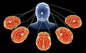
Background imageCentral Nervous System Collection: Conceptual image of multi-brain processing

Conceptual image of multi-brain processing

Background imageCentral Nervous System Collection: Anatomy structure of neurons

Anatomy structure of neurons

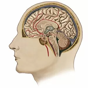
Background imageCentral Nervous System Collection: Cross section of brain with arteries

Cross section of brain with arteries

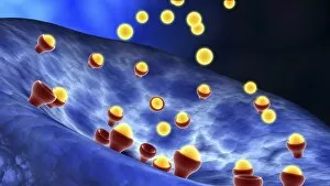
Background imageCentral Nervous System Collection: Conceptual image of synapse receptors

Conceptual image of synapse receptors

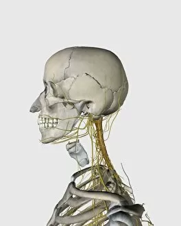
Background imageCentral Nervous System Collection: Medical illustration showing thyroid cartilage and nerves around neck and head area

Medical illustration showing thyroid cartilage and nerves around neck and head area
Medical illustration showing thyroid cartilage and nerves around human neck and head area

Background imageCentral Nervous System Collection: Schematic of the hypothalamus receiving nerve impulses from the body

Schematic of the hypothalamus receiving nerve impulses from the body and sending messages to the circulatory and nervous system

Background imageCentral Nervous System Collection: Human brain showing the layout of the sensory cortex

Human brain showing the layout of the sensory cortex
Coronal section through human brain showing the layout of the sensory cortex

Background imageCentral Nervous System Collection: Conceptual image of synaptic transmission

Conceptual image of synaptic transmission

Background imageCentral Nervous System Collection: Microscopic view of a bipolar neuron

Microscopic view of a bipolar neuron. A bipolar cell is a type of neuron which has two extensions. Bipolar cells are specialized sensory neurons for the transmission of special senses

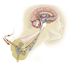
Background imageCentral Nervous System Collection: Endorphins released in the hypothalamus travel down the spinal cord

Endorphins released in the hypothalamus travel down the spinal cord
Serotonin released in the brain travels down the spinal cord to close the pain gates and block pain messages

Background imageCentral Nervous System Collection: Sagittal view of human brain showing the corpus callosum

Sagittal view of human brain showing the corpus callosum

Background imageCentral Nervous System Collection: Human brain with nerves

Human brain with nerves

Background imageCentral Nervous System Collection: Illustration of muscle contraction (with labels)

Illustration of muscle contraction (with labels)

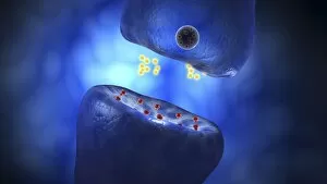
Background imageCentral Nervous System Collection: Close-up view of the synapse in the nervous system

Close-up view of the synapse in the nervous system. A synapse is a structure that permits a neuron (or nerve cell) to pass an electrical or chemical signal to another cell

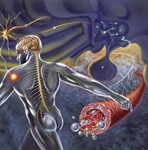
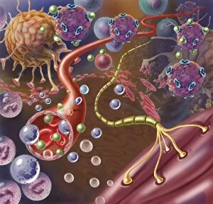
Background imageCentral Nervous System Collection: Nerve with myelin sheath, seen in lower right, connects with muscle

Nerve with myelin sheath, seen in lower right, connects with muscle. Blood vessel and immune cells are seen in the center and upper right of image
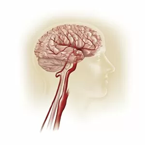
Background imageCentral Nervous System Collection: Side view of human brain showing internal carotid artery

Side view of human brain showing internal carotid artery

Background imageCentral Nervous System Collection: Conceptual image of GABA receptors

Conceptual image of GABA receptors. The GABA receptors are a class of receptors that respond to the neurotransmitter gamma-aminobutyric acid
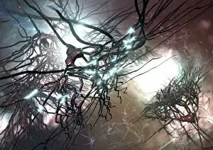
Background imageCentral Nervous System Collection: Conceptual image of a neuron ligntning signal passing

Conceptual image of a neuron ligntning signal passing

Background imageCentral Nervous System Collection: X-ray view of human face, profile view

X-ray view of human face, profile view

Background imageCentral Nervous System Collection: Neurofibromatosis, a genetic disorder of the nervous system

Neurofibromatosis, a genetic disorder of the nervous system
A genetic disorder of the nervous system, neurofibromatosis causes tumors to form on nerves throughout the body, including a type of tumor called an optic nerve glioma that can result in childhood

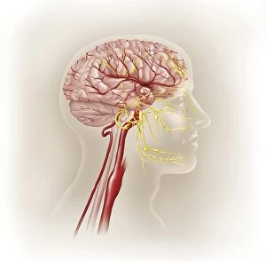
Background imageCentral Nervous System Collection: Detail of ateries of the human head and the trigeminal nerve

Detail of ateries of the human head and the trigeminal nerve

Background imageCentral Nervous System Collection: Microscopic view of inner nervous system

Microscopic view of inner nervous system

Background imageCentral Nervous System Collection: Microscopic view of a multipolar neuron

Microscopic view of a multipolar neuron. Multipolar neurons possess a single axon and many dendrites

Background imageCentral Nervous System Collection: Illustration of action potential of a nerve cell

Illustration of action potential of a nerve cell

Background imageCentral Nervous System Collection: Conceptual image of a neuron

Conceptual image of a neuron

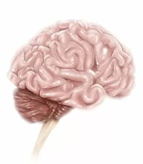
Background imageCentral Nervous System Collection: Human brain anatomy, lateral view, with labels

Human brain anatomy, lateral view, with labels

Background imageCentral Nervous System Collection: Illustration showing cross section of human brain

Illustration showing cross section of human brain
Medicine: encephalon

Background imageCentral Nervous System Collection: Prion disease treatment, artwork C018 / 1838

Prion disease treatment, artwork C018 / 1838
Prion disease treatment. Artwork of three pyramidal neurons (nerve cells). The cell at left is healthy. The cell at back has died because of a prion (pink) infection

Background imageCentral Nervous System Collection: Pyramidal nerve cells, artwork C017 / 2273

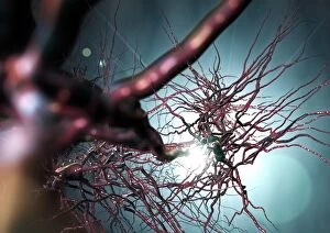
Pyramidal nerve cells, artwork C017 / 2273
Pyramidal nerve cells. Computer artwork of pyramidal nerve cells from the cerebral cortex of the brain. Pyramidal cells are so named for their triangular cell bodies

Background imageCentral Nervous System Collection: Nerve synapse, artwork C017 / 3427

Nerve synapse, artwork C017 / 3427
Nerve synapse. Computer artwork of of a junction, or synapse, between two nerve cells (neurons). As the electrical signal reaches the presynaptic end of a neuron it triggers the release of

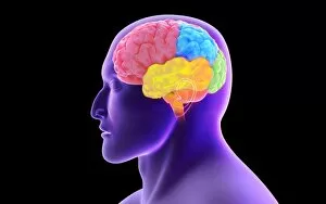
Background imageCentral Nervous System Collection: Brain scan, conceptual image C017 / 7392

Brain scan, conceptual image C017 / 7392
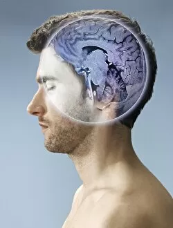
MODEL RELEASED. Brain scan, conceptual composite image

Background imageCentral Nervous System Collection: Brain scan, conceptual image C017 / 7394

Brain scan, conceptual image C017 / 7394
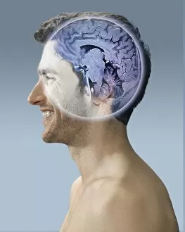
MODEL RELEASED. Brain scan, conceptual composite image

Background imageCentral Nervous System Collection: Brain scan, conceptual image C017 / 7398

Brain scan, conceptual image C017 / 7398
MODEL RELEASED. Brain scan, conceptual composite image

Background imageCentral Nervous System Collection: Brain scan, conceptual image C017 / 7397

Brain scan, conceptual image C017 / 7397
MODEL RELEASED. Brain scan, conceptual composite image

Background imageCentral Nervous System Collection: Nerve synapse, artwork C017 / 3428

Nerve synapse, artwork C017 / 3428
Nerve synapse. Computer artwork of of a junction, or synapse, between two nerve cells (neurons). As the electrical signal reaches the presynaptic end of a neuron it triggers the release of

Background imageCentral Nervous System Collection: Corpus callosum, DTI MRI scan C017 / 7044

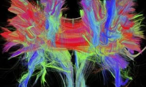
Corpus callosum, DTI MRI scan C017 / 7044
Corpus callosum. Coronal 3D diffusion tensor imaging (DTI) magnetic resonance imaging (MRI) scan of nerve pathways (coloured) in and around the brains corpus callosum

Background imageCentral Nervous System Collection: Pyramidal nerve cells, artwork C017 / 2274

Pyramidal nerve cells, artwork C017 / 2274
Pyramidal nerve cells. Computer artwork of pyramidal nerve cells from the cerebral cortex of the brain. Pyramidal cells are so named for their triangular cell bodies

Background imageCentral Nervous System Collection: Brain scan, conceptual image C017 / 7391

Brain scan, conceptual image C017 / 7391
MODEL RELEASED. Brain scan, conceptual composite image

Background imageCentral Nervous System Collection: Cyclist, artwork F006 / 7385

Cyclist, artwork F006 / 7385
Cyclist, computer artwork

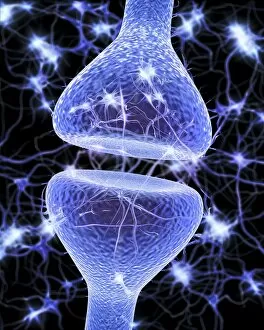
Background imageCentral Nervous System Collection: Nerve synapse, artwork F006 / 7073

Nerve synapse, artwork F006 / 7073
Nerve synapse. Computer artwork of a junction, or synapse, between two nerve cells (neurons, blue). As the electrical signal reaches the presynaptic end of a neuron it triggers the release of

Background imageCentral Nervous System Collection: Baseball player, artwork F006 / 7315

Baseball player, artwork F006 / 7315
Baseball player, computer artwork

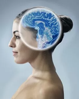
Background imageCentral Nervous System Collection: MRI brain scan and nerve cells F006 / 3595

MRI brain scan and nerve cells F006 / 3595
Computer artwork of a sagittal (side) view of a human head. The largest part of the brain is the cerebrum (upper part). This surrounds the limbic system (curved structures at upper centre)

Background imageCentral Nervous System Collection: Brain and nerve cells, neural network F006 / 3585

Brain and nerve cells, neural network F006 / 3585
Computer artwork of a frontal view of a human brain. In the background a neural network of nerve cells firing

Background imageCentral Nervous System Collection: Nerve synapse, artwork F006 / 7074

Nerve synapse, artwork F006 / 7074
Nerve synapse. Computer artwork of a junction, or synapse, between two nerve cells (neurons, blue). As the electrical signal reaches the presynaptic end of a neuron it triggers the release of

Background imageCentral Nervous System Collection: Human brain, artwork F006 / 6313

Human brain, artwork F006 / 6313
Human brain, computer artwork. The front of the brain is at bottom

Background imageCentral Nervous System Collection: Cyclist, artwork F006 / 7388

Cyclist, artwork F006 / 7388
Cyclist, computer artwork

Background imageCentral Nervous System Collection: Sleeping man, artwork F006 / 7362

Sleeping man, artwork F006 / 7362
Sleeping man, computer artwork

Background imageCentral Nervous System Collection: Nerve synapse, artwork F006 / 7090

Nerve synapse, artwork F006 / 7090
Nerve synapse. Computer artwork of a junction, or synapse, between two nerve cells (neurons, blue). As the electrical signal reaches the presynaptic end of a neuron it triggers the release of


All Professionally Made to Order for Quick Shipping

The central nervous system, the intricate network that governs our every thought and movement, is a marvel of complexity. From the delicate cerebellum tissue to the detailed light micrograph capturing its essence, we are reminded of its importance in maintaining balance and coordination. Anatomy comes alive as we explore the human brain from an inferior view. The intricacy of brain fibers is revealed through DTI MRI scans like C017/7099 and C017/7035, showcasing their vital role in transmitting information throughout this extraordinary organ. Artistic renderings bring us closer to understanding the medulla oblongata's significance within the brain. Its portrayal in various artworks allows us to appreciate how it controls essential functions such as breathing and heart rate. As we delve deeper into studying the central nervous system, models of the human brain provide invaluable insights into its structure and organization. Lateral views reveal countless regions responsible for cognition, emotion regulation, sensory perception, and motor control. Microscope slides offer glimpses into nerve cells' intricate architecture—a testament to their ability to transmit electrical signals at lightning speed. Meanwhile, glial stem cell cultures captured under a light microscope remind us of their crucial role in supporting neuronal function. Finally, artistic representations unveil the limbic system's enigmatic nature—an interconnected web responsible for emotions and memory formation. These captivating artworks allow us to visualize this complex network within our brains. Exploring these hints provides a glimpse into the awe-inspiring world of our central nervous system—the very foundation upon which our thoughts, actions, memories reside—reminding us just how remarkable our brains truly are.