Fraud Blocker Skip to main content

Biochemical Collection (page 2)

"Unveiling the Intricacies of Biochemical World: From Anaesthetic Inhibiting Ion Channels to DNA Discoveries" Delving into the depths wonders

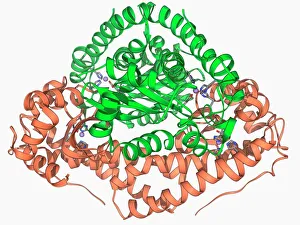
Background imageBiochemical Collection: Manganese superoxide dismutase enzyme F006 / 9423

Manganese superoxide dismutase enzyme F006 / 9423
Manganese superoxide dismutase enzyme, molecular model. This enzyme scavenges and decomposes the potentially toxic first reduction product, superoxide, of aerobic respiration

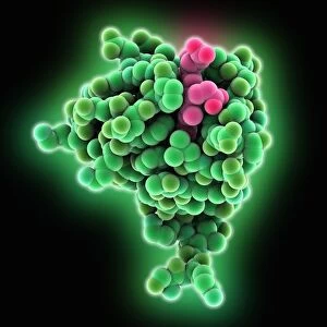
Background imageBiochemical Collection: SARS coronavirus protein

SARS coronavirus protein. Molecular model of the ORF-9b protein produced by the SARS (severe acute respiratory syndrome) coronavirus

Background imageBiochemical Collection: Cytochrome b5 molecule C015 / 6696

Cytochrome b5 molecule C015 / 6696
Cytochrome b5. Molecular model of cytochrome b5 from a cows liver. Cytochrome molecules perform oxidation and reduction reactions for electron transport

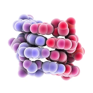
Background imageBiochemical Collection: Z-DNA tetramer molecule C015 / 6557

Z-DNA tetramer molecule C015 / 6557
Z-DNA (deoxyribonucleic acid) tetramer, molecular model. DNA is composed of two strands twisted into a double helix. This is a tetramer of the molecule, containing four strands

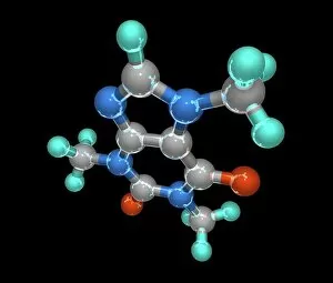
Background imageBiochemical Collection: Caffeine, molecular model

Caffeine, molecular model. Atoms are represented as spheres and are colour-coded: carbon (grey), hydrogen (green), oxygen (red) and nitrogen (blue)

Background imageBiochemical Collection: Shared DNA in humans and chimps, art

Shared DNA in humans and chimps, art
Shared DNA between humans and chimps, conceptual artwork. The humans look surprised to see the chimp so close to them in the double helix of DNA (deoxyribonucleic acid)

Background imageBiochemical Collection: Mitochondrial DNA

Mitochondrial DNA. Computer artwork of the genetic material (DNA, deoxyribonucleic acid) found in the cell structures called mitochondria

Background imageBiochemical Collection: Vinegar production, 19th century

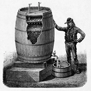
Vinegar production, 19th century cutaway artwork. This is a trickling method developed in 1823 by the German chemist Schutzenbach. The barrel is divided into three sections

Background imageBiochemical Collection: Collagen synthesis and assembly, artwork

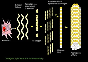
Collagen synthesis and assembly, artwork. At left is a fibroblast, the cell that synthesises helical protein chains of collagen (wavy lines)

Background imageBiochemical Collection: Nerve damage and stem cells, artwork

Nerve damage and stem cells, artwork
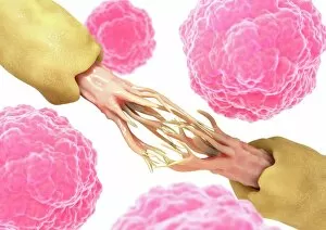
Nerve damage and stem cells, computer artwork. Stem cells are undifferentiated cells that can produce other types of cell when they divide

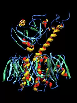
Background imageBiochemical Collection: Cholera toxin, molecular model

Cholera toxin, molecular model
Cholera toxin. Molecular model of the secondary structure of cholera enterotoxin (intestinal toxin). The molecule consists of two subunits, A (top) and B (bottom)

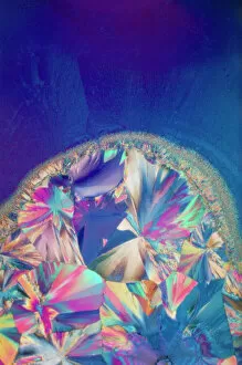
Background imageBiochemical Collection: Progesterone hormone

Progesterone hormone
Polarised light micrograph of crystals of progesterone. X 40

Background imageBiochemical Collection: Isotretinoin anti-acne drug

Isotretinoin anti-acne drug, molecular model. Atoms are represented as tubes and are colour- coded; carbon (yellow), hydrogen (white) and oxygen (red)

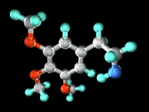
Background imageBiochemical Collection: Mescaline hallucinogenic drug molecule

Mescaline hallucinogenic drug molecule
Mescaline hallucinogenic drug, molecular model. Mescaline is a hallucinogenic drug, produced from the dried tops (buttons) of the peyote cactus (Lophophora williamsii)

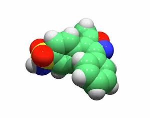
Background imageBiochemical Collection: Valdecoxib anti-inflammatory drug

Valdecoxib anti-inflammatory drug
Valdecoxib, computer model. This drug was used in the treatment of osteoarthritis, rheumatoid arthritis and menstrual symptoms under the trade name Bextra

Background imageBiochemical Collection: Paclitaxel drug molecule

Paclitaxel drug molecule
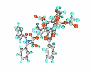
Paclitaxel. Computer model of a molecule of the drug paclitaxel. It is sold under the brand name Taxol. It is a chemotherapy drug, used to treat cancers

Background imageBiochemical Collection: Human chromosomes, SEM C013 / 5002

Human chromosomes, SEM C013 / 5002
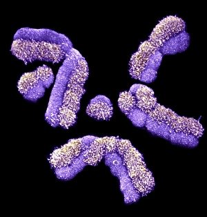
Human chromosomes. Coloured scanning electron micrograph (SEM) of human chromosomes prepared with the harlequin staining technique

Background imageBiochemical Collection: DNA strands, illustration

DNA strands, illustration
DNA strands. Computer illustration showing the structure of double stranded DNA (deoxyribonucleic acid) molecules. DNA is composed of two strands twisted into a double helix

Background imageBiochemical Collection: International biohazard symbol

International biohazard symbol warning of a potential biological hazard. This symbol is used worldwide in laboratories and on containers where biologically active agents are present

Background imageBiochemical Collection: Argonaute protein molecule F006 / 9526

Argonaute protein molecule F006 / 9526
Argonaute protein, molecular model. This protein forms the RNA-induced silencing complex (RISC) along with a small interfering RNA (ribonucleic acid) molecule

Background imageBiochemical Collection: TFAM transcription factor bound to DNA C015 / 7059

TFAM transcription factor bound to DNA C015 / 7059
TFAM transcription factor bound to DNA, molecular model. Human mitochondrial transcription factor A (TFAM, green) bound to a strand of DNA (deoxyribonucleic acid, blue and pink)

Background imageBiochemical Collection: Insulin production plant C017 / 9435

Insulin production plant C017 / 9435
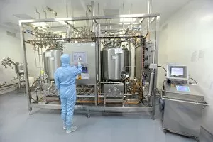
Insulin production plant. Worker in the reactor room of a factory producing genetically engineered insulin. Photographed at the Novouralsk Medsintez Plant, Novouralsk, Sverdlov Oblast, Russia

Background imageBiochemical Collection: Vitamin B12 injection, conceptual artwork

Vitamin B12 injection, conceptual artwork
Vitamin B12 injection, conceptual composite artwork

Background imageBiochemical Collection: DNA by tunnelling microscope

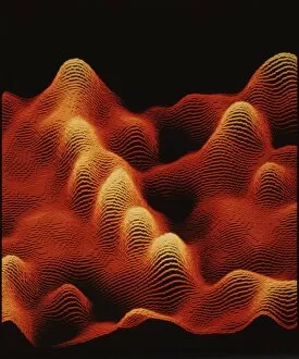
DNA by tunnelling microscope
False-colour scanning tunnelling micrograph (STM) of DNA. A sample of uncoated, double-stranded DNA was dissolved in a salt solution & deposited on graphite prior to being imaged in air by the STM

Background imageBiochemical Collection: DNA structure, artwork C017 / 7218

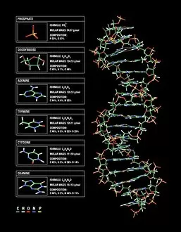
DNA structure, artwork C017 / 7218
DNA structure. Computer artwork showing the structure of a double stranded DNA (deoxyribonucleic acid) molecule (right) and its components (left)

Background imageBiochemical Collection: Argonaute protein and microRNA F006 / 9752

Argonaute protein and microRNA F006 / 9752
Argonaute protein. Molecular model of human argonaute-2 protein complexed with microRNA (micro ribonucleic acid). This protein is part of the RNA-induced silencing complex (RISC)

Background imageBiochemical Collection: Immunoglobulin G antibody and egg white F006 / 9682

Immunoglobulin G antibody and egg white F006 / 9682
Immunoglobulin G and egg white. Molecular model of an immunoglobulin G (IgG) antibody bound to a molecule of egg white. This is the most abundant immunoglobulin and is found in all body fluids

Background imageBiochemical Collection: Cytochrome P450 complex F006 / 9669

Cytochrome P450 complex F006 / 9669
Cytochrome P450 complex. Molecular model of a complex composed of cytochrome P450, carbon monoxide and camphor. Cytochrome molecules perform oxidation and reduction reactions for electron transport

Background imageBiochemical Collection: Succinyl-CoA synthetase enzyme F006 / 9592

Succinyl-CoA synthetase enzyme F006 / 9592
Succinyl-CoA synthetase bound to GTP, molecular model. Also known as succinyl coenzyme A synthetase (SCS), this enzyme catalyses the reversible reaction between succinyl-CoA and succinic acid

Background imageBiochemical Collection: RNA-induced silencing complex F006 / 9586

RNA-induced silencing complex F006 / 9586
RNA-induced silencing complex (RISC), molecular model. This complex consists of a bacterial argonaute protein (top) bound to a small interfering RNA (siRNA) molecule (red and blue)

Background imageBiochemical Collection: Foot-and-mouth disease virus F006 / 9556

Foot-and-mouth disease virus F006 / 9556
Foot-and-mouth disease virus. Molecular model of the foot-and-mouth disease (FMD) virus (Aphtae epizooticae) protein coat (capsid)

Background imageBiochemical Collection: Adenovirus penton base protein F006 / 9542

Adenovirus penton base protein F006 / 9542
Adenovirus penton base protein, molecular model. This protein molecule is a subunit called a penton, forming the vertices of the capsid of this adenovirus

Background imageBiochemical Collection: Rhinovirus 16 capsid, molecular model F006 / 9431

Rhinovirus 16 capsid, molecular model F006 / 9431
Rhinovirus 16 capsid, molecular model. This is human rhinovirus 16. The rhinovirus infects the upper respiratory tract and is the cause of the common cold. It is spread by coughs and sneezes

Background imageBiochemical Collection: Citrate acid cycle enzyme F006 / 9305

Citrate acid cycle enzyme F006 / 9305
Citrate acid cycle enzyme. Molecular model of the enzyme dihydrolipoamide succinyltransferase. This enzyme is involved in the citric acid (or Krebs) cycle

Background imageBiochemical Collection: Pyruvate dehydrogenase complex C018 / 9192

Pyruvate dehydrogenase complex C018 / 9192
Pyruvate dehydrogenase complex, 3D model. This enzyme complex is responsible for the step that links glycolysis to the citric acid (Krebs) cycle

Background imageBiochemical Collection: High-contrast direct DNA image, TEM

High-contrast direct DNA image, TEM
High-contrast direct DNA image. Coloured transmission electron micrograph (TEM) of the first high-contrast direct image of a bundle (fibre) of strands of DNA (deoxyribonucleic acid)

Background imageBiochemical Collection: Cell membrane ion channels, artwork C016 / 7689

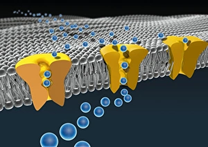
Cell membrane ion channels, artwork C016 / 7689
Cell membrane ion channels. Computer artwork of a section through the membrane of an animal cell, showing transmembrane ion channel proteins (yellow)

Background imageBiochemical Collection: Valproic acid anticonvulsant molecule C014 / 2296

Valproic acid anticonvulsant molecule C014 / 2296
Valproic acid. Molecular model of the anticonvulsant and mood-stabilising drug valproic acid. It is used to treat epilepsy, seizures, bipolar disorder and depression

Background imageBiochemical Collection: MscL ion channel protein structure

MscL ion channel protein structure. Molecular model showing the protein structure of a Mechanosensitive Channel of Large Conductance (MscL) from a Mycobacterium tuberculosis bacterium

Background imageBiochemical Collection: Immunoglobulin A, molecular model

Immunoglobulin A, molecular model
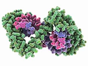
Immunoglobulin A. Molecular model of the structure of the antibody immunoglobulin A (IgA). This is the secretory dimeric form (sIgA), the main immunoglobulin found in secretions such as saliva

Background imageBiochemical Collection: Effects of ecstasy on brain function

Effects of ecstasy on brain function. Computer artwork representing the effects of MDMA (3, 4-methylenedioxy-N-methylamphetamine), or ecstasy, on the nerves (centre) and synapses (nerve junctions)

Background imageBiochemical Collection: Adenovirus hexon protein

Adenovirus hexon protein, molecular model. Hexon proteins are part of the protein coat or shell (capsid) of adenoviruses. In viruses

Background imageBiochemical Collection: Immunoglobulin G antibody molecule C016 / 4462

Immunoglobulin G antibody molecule C016 / 4462
Immunoglobulin G antibody molecule. Computer artwork of a model of the secondary structure of immunoglobulin G (IgG). This is the most abundant immunoglobulin and is found in all body fluids

Background imageBiochemical Collection: X-ray crystallography C016 / 3824

X-ray crystallography C016 / 3824
X-ray crystallography. Researcher using an X-ray machine to obtain crystal diffraction patterns of proteins for 3-D imaging of enzymes

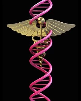
Background imageBiochemical Collection: Caduceus with DNA, artwork C013 / 9990

Caduceus with DNA, artwork C013 / 9990
Caduceus with DNA. Computer artwork of the Caduceus symbol entwined by a strand of DNA (deoxyribonucleic acid). The caduceus is the traditional symbol of the Greek god Hermes

Background imageBiochemical Collection: Zebra fish embryo, SEM C013 / 9587

Zebra fish embryo, SEM C013 / 9587
Zebra fish embryo. Coloured scanning electron micrograph (SEM) of the embryo of a zebrafish (Danio rerio), showing its eyes either side of its large open mouth (centre)

Background imageBiochemical Collection: Coagulation factor complex molecule C014 / 0139

Coagulation factor complex molecule C014 / 0139
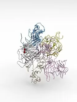
Coagulation factor complex molecule. Molecular model showing the interaction between coagulation factor VIII (FVIII, pink, blue and yellow) and factor IXa (FIXa, cream and grey)

Background imageBiochemical Collection: Creation of oil using designer microbes

Creation of oil using designer microbes. Conceptual computer artwork depicting an oil well pump in a petri dish, representing the use of artificially created micro-organisms to produce oil


All Professionally Made to Order for Quick Shipping

"Unveiling the Intricacies of Biochemical World: From Anaesthetic Inhibiting Ion Channels to DNA Discoveries" Delving into the depths wonders, scientists have uncovered an anaesthetic that inhibits an ion channel (C015 / 6718), shedding light on new possibilities for pain management. The enigmatic double-stranded RNA molecule reveals its secrets, captivating researchers with its role in gene regulation and potential therapeutic applications. Peering into the intricate world of DNA transcription through a molecular model, scientists unravel the mysteries behind genetic information transfer and cellular function. Captured under a microscope's gaze, caffeine crystals dazzle with their vibrant beauty, reminding us of this ubiquitous stimulant's impact on our daily lives. The iconic DNA molecule stands tall as a symbol of life's blueprint, holding within it the key to our genetic heritage and evolutionary history. Immunoglobulin G antibody molecule emerges as a formidable defender against pathogens, showcasing nature's ingenious immune system at work. Through mesmerizing crystal formations seen under intense magnification, EDTA crystals reveal their significance in chelation therapy and metal ion sequestration processes. Oxytocin hormone crystals shimmer like precious gems when observed through polarized light microscopy (PLM C016 / 7196), highlighting its crucial role in social bonding and reproductive functions. Watson and Crick forever etched their names in scientific history by unravelling the structure of DNA; their groundbreaking discovery paved the way for countless advancements in genetics research. Celebrated author Isaac Asimov not only captivated readers with his science fiction tales but also left an indelible mark as a biochemist who popularized complex scientific concepts for all to comprehend and appreciate. Artistic renditions bring metabolic enzymes to life as they orchestrate vital chemical reactions within cells – true catalysts that drive life's intricate processes.