Fraud Blocker Skip to main content

Biochemical Collection (page 8)

"Unveiling the Intricacies of Biochemical World: From Anaesthetic Inhibiting Ion Channels to DNA Discoveries" Delving into the depths wonders

Background imageBiochemical Collection: Ricin A-chain, artwork C017 / 3654

Ricin A-chain, artwork C017 / 3654
Ricin A-chain. Computer artwork showing the enzymatically active A-chain from a molecule of the toxic protein ricin. Ricin comprises two entwined amino acid chains; A (seen here) and B (not shown)

Background imageBiochemical Collection: Sirtuin enzyme and p53, artwork C017 / 3660

Sirtuin enzyme and p53, artwork C017 / 3660
Sirtuin enzyme and p53. Computer artwork of a sirtuin (Sir2) enzyme (blue) bound to a p53 peptide (pink). Sir2 enzymes form a unique class of NAD(+)

Background imageBiochemical Collection: EcoRV restriction enzyme molecule C014 / 2114

EcoRV restriction enzyme molecule C014 / 2114
EcoRV restriction enzyme. Molecular model of the type II restriction enzyme EcoRV (white and gold) bound to a cleaved section of DNA (deoxyribonucleic acid, orange and yellow)

Background imageBiochemical Collection: Genetic code, artwork F006 / 8998

Genetic code, artwork F006 / 8998
Genetic code, computer artwork

Background imageBiochemical Collection: EcoRV restriction enzyme molecule C014 / 2116

EcoRV restriction enzyme molecule C014 / 2116
EcoRV restriction enzyme. Molecular model of the type II restriction enzyme EcoRV (purple and blue) bound to a DNA molecule (deoxyribonucleic acid, pink and white)

Background imageBiochemical Collection: Genetic code, artwork F006 / 8997

Genetic code, artwork F006 / 8997
Genetic code, computer artwork

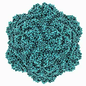
Background imageBiochemical Collection: Tobacco necrosis virus capsid

Tobacco necrosis virus capsid, molecular model. This plant virus infects a wide rage of plants, including the tobacco plant for which it is named. The virus causes tissue death (necrosis)

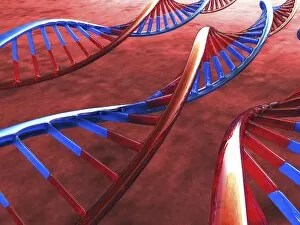
Background imageBiochemical Collection: DNA molecule, artwork F006 / 8969

DNA molecule, artwork F006 / 8969
DNA molecule, computer artwork

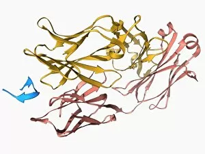
Background imageBiochemical Collection: Ricin molecule, artwork C017 / 3649

Ricin molecule, artwork C017 / 3649
Ricin molecule. Computer artwork showing the structure of a molecule of the toxic protein ricin. Ricin comprises two entwined amino acid chains; A (yellow) and B (blue)

Background imageBiochemical Collection: Insulin molecule C014 / 2121

Insulin molecule C014 / 2121
Insulin molecule. Molecular module of insulin showing its secondary structure. Insulin is a hormone produced by the pancreas

Background imageBiochemical Collection: TATA box-binding protein complex C017 / 7083

TATA box-binding protein complex C017 / 7083
TATA box-binding protein complex. Molecular model showing a TATA box-binding protein (TBP, green) complexed with a strand of DNA (deoxyribonucleic acid, yellow) and transcription factor IIB

Background imageBiochemical Collection: DNA molecules, artwork F006 / 8775

DNA molecules, artwork F006 / 8775
DNA molecules, computer artwork

Background imageBiochemical Collection: DNA molecules, artwork F006 / 8776

DNA molecules, artwork F006 / 8776
DNA molecules, computer artwork

Background imageBiochemical Collection: Genetic engineering, conceptual artwork F006 / 8780

Genetic engineering, conceptual artwork F006 / 8780
Genetic engineering, conceptual computer artwork

Background imageBiochemical Collection: DNA molecules, artwork F006 / 8774

DNA molecules, artwork F006 / 8774
DNA molecules, computer artwork

Background imageBiochemical Collection: EcoRV restriction enzyme molecule C014 / 2115

EcoRV restriction enzyme molecule C014 / 2115
EcoRV restriction enzyme. Molecular model of the type II restriction enzyme EcoRV (purple and blue) bound to a DNA molecule (deoxyribonucleic acid, pink and white)

Background imageBiochemical Collection: Palmitoyloleoyl PE lipid bilayer F006 / 9780

Palmitoyloleoyl PE lipid bilayer F006 / 9780
Palmitoyloleoyl PE (POPE) lipid bilayer, computer simulation. This is a model of the phospholipid bilayer of the inner bacterial membrane

Background imageBiochemical Collection: Rubisco enzyme molecule F006 / 9776

Rubisco enzyme molecule F006 / 9776
Rubisco. Molecular model of the enzyme rubisco (ribulose bisphosphate carboxylase oxygenase) complexed with 2-carboxyarabinitol biphosphate

Background imageBiochemical Collection: Rubisco enzyme molecule F006 / 9779

Rubisco enzyme molecule F006 / 9779
Rubisco. Molecular model of the enzyme rubisco (ribulose bisphosphate carboxylase oxygenase) complexed with ribulose-1, 5-biphosphate

Background imageBiochemical Collection: NADP-dependent isocitrate dehydrogenase F006 / 9778

NADP-dependent isocitrate dehydrogenase F006 / 9778
NADP-dependent isocitrate dehydrogenase, molecular model. This enzyme catalyses the third step in the citric acid (or Krebs) cycle, the process by which mitochondria convert glucose to energy

Background imageBiochemical Collection: Catalase, molecular model F006 / 9774

Catalase, molecular model F006 / 9774
Catalase. Molecular model of catalase from a cow liver. This enzyme to water and oxygen. Hydrogen peroxide is a highly toxic byproduct of a number of normal cellular processes

Background imageBiochemical Collection: Glycogen phosphorylase molecule F006 / 9775

Glycogen phosphorylase molecule F006 / 9775
Glycogen phosphorylase. Molecular model of glycogen phosphorylase bound to AMP (adenosine monophosphate). This is an enzyme involved in breaking down glycogen

Background imageBiochemical Collection: Triose phosphate isomerase molecule F006 / 9777

Triose phosphate isomerase molecule F006 / 9777
Triose phosphate isomerase (TPI), molecular model. TPI is essential for glycolysis and catalyses the reversible interconversion of dihydroxyacetone phosphate and glyceraldehyde-3-phosphate

Background imageBiochemical Collection: Bacterial alpha-hemolysin toxin F006 / 9771

Bacterial alpha-hemolysin toxin F006 / 9771
Bacterial alpha-hemolysin toxin, molecular model. This toxin is secreted by the bacterium Staphylococcus aureus. It is an example of a pore-forming toxin

Background imageBiochemical Collection: Metabolic enzyme molecule F006 / 9770

Metabolic enzyme molecule F006 / 9770
Metabolic enzyme. Molecular model of the enzyme aconitase with isocitrate bound. Aconitase is involved in the citric acid (or Krebs) cycle

Background imageBiochemical Collection: Dihydrofolate reductase molecule F006 / 9772

Dihydrofolate reductase molecule F006 / 9772
Dihydrofolate reductase, molecular model. This enzyme converts the vitamin folic acid into a coenzyme

Background imageBiochemical Collection: HIV-1 protease and inhibitor F006 / 9773

HIV-1 protease and inhibitor F006 / 9773
HIV-1 protease and inhibitor. Molecular model of the enzyme HIV-1 protease (pink and blue ribbons) bound to an inhibitor molecule (centre)

Background imageBiochemical Collection: Ribonuclease A molecule F006 / 9768

Ribonuclease A molecule F006 / 9768
Ribonuclease A (RNAse A), molecular model. Ribonuclease (RNase) is a type of nuclease that catalyses the degradation of RNA (ribonucleic acid)

Background imageBiochemical Collection: Pepsin stomach enzyme F006 / 9767

Pepsin stomach enzyme F006 / 9767
Pepsin stomach enzyme, molecular model. Pepsin is a protease enzyme that is secreted as part of gastric juice into the stomach in an inactive form known as pepsinogen

Background imageBiochemical Collection: Flock house virus capsid F006 / 9755

Flock house virus capsid F006 / 9755
Flock house virus capsid, molecular model. The flock house virus is a member of the Nodaviridae family. It kills the New Zealand grass grub insect

Background imageBiochemical Collection: Xylose isomerase complex F006 / 9765

Xylose isomerase complex F006 / 9765
Xylose isomerase complex. Molecular model of the enzyme D-xylose isomerase bound to the sugar alcohol sorbitol. D-xylose isomerase is involved in fructose and mannose metabolism

Background imageBiochemical Collection: H-Ras p21 oncogene protein F006 / 9766

H-Ras p21 oncogene protein F006 / 9766
H-Ras p21 oncogene protein, molecular model. The Ras proteins are involved in transmitting signals within cells. Excessive signalling can lead to conditions such as cancer

Background imageBiochemical Collection: Phosphofructokinase bacterial enzyme F006 / 9762

Phosphofructokinase bacterial enzyme F006 / 9762
Phosphofructokinase enzyme, molecular model. This enzyme, from the bacterium Bacillus stearothermophilus, is involved in regulating the process of releasing energy from glucose

Background imageBiochemical Collection: tRNA molecule F006 / 9764

tRNA molecule F006 / 9764
Transfer RNA (tRNA), molecular model. tRNA (transfer ribonucleic acid) translates messenger RNA (mRNA) into a protein product

Background imageBiochemical Collection: H-Ras p21 oncogene protein F006 / 9763

H-Ras p21 oncogene protein F006 / 9763
H-Ras p21 oncogene protein, molecular model. The Ras proteins are involved in transmitting signals within cells. Excessive signalling can lead to conditions such as cancer

Background imageBiochemical Collection: Kinase inhibitor complex F006 / 9760

Kinase inhibitor complex F006 / 9760
Kinase inhibitor complex. Molecular model of a leucettine kinase inhibitor bound to a serine threonine kinase protein

Background imageBiochemical Collection: Marburg viral protein 35 and RNA F006 / 9759

Marburg viral protein 35 and RNA F006 / 9759
Marburg viral protein 35 and RNA. Molecular model of the Marburg viral protein 35 (VP35) bound to a molecule of double stranded RNA (ribonucleic acid)

Background imageBiochemical Collection: Insulin molecule F006 / 9761

Insulin molecule F006 / 9761
Insulin molecule. Molecular model of the hormone insulin from a pig. Insulin consists of two peptide chains, A and B, which are linked by disulphide bridges

Background imageBiochemical Collection: Eye lens protein molecule F006 / 9758

Eye lens protein molecule F006 / 9758
Eye lens protein. Molecular model of gammaB-crystallin, a protein found in the lens of the eye. The regular arrangement of the protein in the lens is thought to be responsible for its transparency

Background imageBiochemical Collection: Methionine aminopeptidase molecule F006 / 9756

Methionine aminopeptidase molecule F006 / 9756
Methionine aminopeptidase, molecular model. This enzyme removes the amino acid methionine from proteins

Background imageBiochemical Collection: Hepatitis C glycoprotein and antibody F006 / 9757

Hepatitis C glycoprotein and antibody F006 / 9757
Hepatitis C glycoprotein and antibody. Molecular model of the E2 envelope glycoprotein from the hepatitis C virus bound to a neutralising antibody

Background imageBiochemical Collection: Malignant brain-tumor-like protein F006 / 9754

Malignant brain-tumor-like protein F006 / 9754
Malignant brain-tumour-like protein. Molecular model of the human lethal(3) malignant brain-tumour-like protein (Malignant brain-tumour-like protein (L3MBTL3)

Background imageBiochemical Collection: UV-damaged DNA-binding protein and DNA F006 / 9750

UV-damaged DNA-binding protein and DNA F006 / 9750
UV-damaged DNA-binding protein and DNA. Molecular model of UV-damaged DNA-binding protein (UV-DDB) complexed with DNA (deoxyribonucleic acid, red and blue)

Background imageBiochemical Collection: Plexin signal transduction molecule F006 / 9751

Plexin signal transduction molecule F006 / 9751
Plexin signal transduction molecule. Molecular model of plexin-A4 a signal transduction protein that is involved in neural maintenance and regeneration

Background imageBiochemical Collection: Multidrug efflux pump molecule F006 / 9748

Multidrug efflux pump molecule F006 / 9748
Multidrug efflux pump. Molecular model of the multidrug efflux pump AcrB from the bacterium Escherichia coli transporting two doxorubicin molecules

Background imageBiochemical Collection: RNA triplet repeat expansion F006 / 9749

RNA triplet repeat expansion F006 / 9749
RNA triplet repeat expansion. Molecular model of a CUG triplet repeat expansion in a molecule of double stranded RNA (ribonucleic acid)

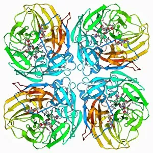
Background imageBiochemical Collection: Flu virus surface protein and drug F006 / 9745

Flu virus surface protein and drug F006 / 9745
Flu virus surface protein and drug. Molecular model of the neuraminidase glycoprotein enzyme from on the surface of the influenza A (flu) virus bound to the drug zanamivir

Background imageBiochemical Collection: Human lysine-specific demethylase F006 / 9747

Human lysine-specific demethylase F006 / 9747
Human lysine-specific demethylase, molecular model. This enzyme removes methyl groups from lysine residues in histones (proteins that package DNA)


All Professionally Made to Order for Quick Shipping

"Unveiling the Intricacies of Biochemical World: From Anaesthetic Inhibiting Ion Channels to DNA Discoveries" Delving into the depths wonders, scientists have uncovered an anaesthetic that inhibits an ion channel (C015 / 6718), shedding light on new possibilities for pain management. The enigmatic double-stranded RNA molecule reveals its secrets, captivating researchers with its role in gene regulation and potential therapeutic applications. Peering into the intricate world of DNA transcription through a molecular model, scientists unravel the mysteries behind genetic information transfer and cellular function. Captured under a microscope's gaze, caffeine crystals dazzle with their vibrant beauty, reminding us of this ubiquitous stimulant's impact on our daily lives. The iconic DNA molecule stands tall as a symbol of life's blueprint, holding within it the key to our genetic heritage and evolutionary history. Immunoglobulin G antibody molecule emerges as a formidable defender against pathogens, showcasing nature's ingenious immune system at work. Through mesmerizing crystal formations seen under intense magnification, EDTA crystals reveal their significance in chelation therapy and metal ion sequestration processes. Oxytocin hormone crystals shimmer like precious gems when observed through polarized light microscopy (PLM C016 / 7196), highlighting its crucial role in social bonding and reproductive functions. Watson and Crick forever etched their names in scientific history by unravelling the structure of DNA; their groundbreaking discovery paved the way for countless advancements in genetics research. Celebrated author Isaac Asimov not only captivated readers with his science fiction tales but also left an indelible mark as a biochemist who popularized complex scientific concepts for all to comprehend and appreciate. Artistic renditions bring metabolic enzymes to life as they orchestrate vital chemical reactions within cells – true catalysts that drive life's intricate processes.