Framed Print > Animals > Mammals > Muridae > House Mouse
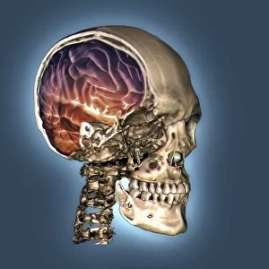
Framed Print : Human skull, X-ray


![]()
Framed Photos from Science Photo Library
Human skull, X-ray
Human skull. Coloured X-ray of a sagittal section through a human skull. The skull has been sliced in half down the centre, revealing details of the internal structures. These include the delicate bones and spaces of the paranasal sinuses (around the nose and eyes, centre left). The fused bones of the cranium encase and protect the brain. The nasal, cheek and upper jaw bones are fused to the cranium to form the facial skeleton (left). The lower jaw bone (mandible, lower left), is attached at flexible joints, allowing it to move. The upper and lower jaw bones contain 32 permanent teeth (lower left). The eye sockets (one at far left) house the eyes
Science Photo Library features Science and Medical images including photos and illustrations
Media ID 6420133
© D. ROBERTS/SCIENCE PHOTO LIBRARY
Bone Bones Bones Cranium Eye Socket Half Halved Jaw Bone Jaws Lower Mandible People Person Persons Profile Radiograph Radiography Sagittal Skeletal Teeth Tooth Upper Section Sectioned
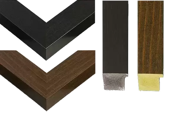
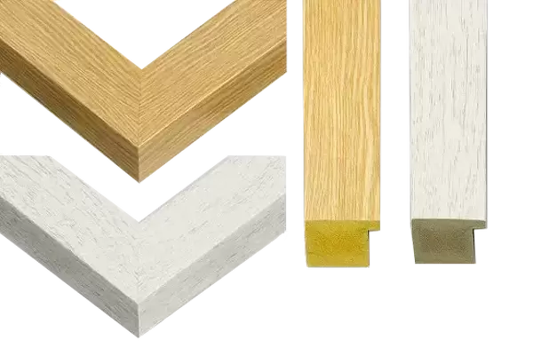
30"x26" (78x68cm) Modern Frame
Discover the intricacies of human anatomy with our captivating Framed Prints from Media Storehouse, featuring the mesmerizing "Human skull, X-ray" image by Science Photo Library. This striking X-ray depicts a sagittal section through a human skull, revealing the intricate details of its internal structures in vibrant colors. Hang this scientific masterpiece in your home or office to ignite conversations and inspire a deeper appreciation for the complexities of the human body. Our high-quality frames enhance the image, ensuring a stunning presentation that is sure to impress. Order yours today and bring the wonder of science into your space.
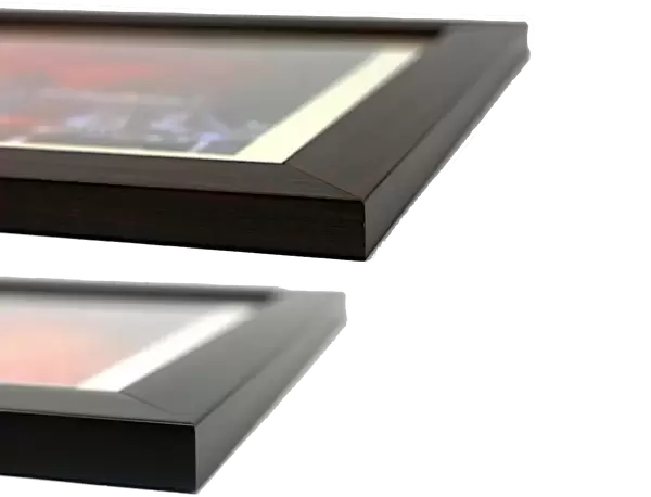
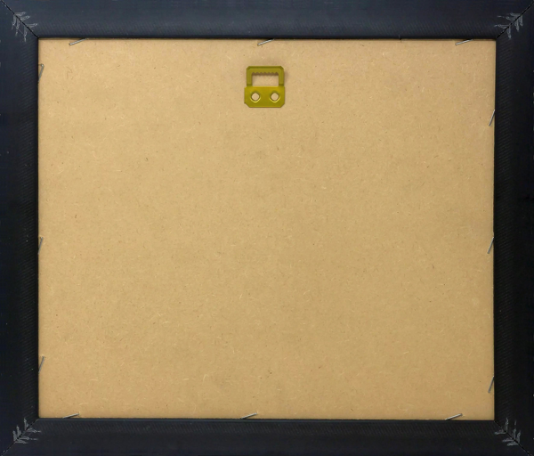
Wood effect frame, card mounted, 24x20 archival quality photo print. Overall outside dimensions 30x26 inches (76x68cm). Environmentally and ozone friendly, 43mm wide x 32mm Polycore® moulding has the look of real wood, is durable and light and easy to hang. Biodegradable and made with non-chlorinated gases (no toxic fumes) it is efficient; producing 100 tons of polystyrene can save 300 tons of trees! Prints are glazed with lightweight, shatterproof, optical clarity acrylic (providing the same general protection from the environment as glass). The back is stapled hardboard with a sawtooth hanger attached. Note: To minimise original artwork cropping, for optimum layout, and to ensure print is secure, the visible print may be marginally smaller
Contemporary Framed and Mounted Prints - Professionally Made and Ready to Hang
Estimated Image Size (if not cropped) is 59.9cm x 58.9cm (23.6" x 23.2")
Estimated Product Size is 78.2cm x 68.2cm (30.8" x 26.9")
These are individually made so all sizes are approximate
Artwork printed orientated as per the preview above, with landscape (horizontal) orientation to match the source image.
FEATURES IN THESE COLLECTIONS
> Animals
> Mammals
> Muridae
> House Mouse
> Science Photo Library
> Specialist Imaging
EDITORS COMMENTS
This print showcases the intricate details of a human skull through a coloured X-ray. The sagittal section reveals the internal structures, providing an extraordinary glimpse into our anatomy. As we explore this image, we can observe the delicate bones and spaces of the paranasal sinuses surrounding the nose and eyes at the center left. The cranium, composed of fused bones, acts as a protective encasement for our brain. Moving towards the left side of this halved skull, we encounter the facial skeleton formed by fusion between nasal, cheek, and upper jaw bones with the cranium. This integration creates a sturdy foundation for our face's structure. On further examination, we notice that flexible joints attach to the lower jaw bone or mandible on its lower left side—allowing us to move it effortlessly. The upper and lower jaws contain 32 permanent teeth—a testament to their vital role in chewing and digestion—displayed prominently on their lower left portion. Additionally, nestled within eye sockets located at far-left lies another fascinating aspect—the housing for our precious eyes. Through this radiograph's lens-like perspective, Science Photo Library has beautifully captured both artistry and science in one frame—an awe-inspiring reminder of how intricately designed our bodies are.
MADE IN THE UK
Safe Shipping with 30 Day Money Back Guarantee
FREE PERSONALISATION*
We are proud to offer a range of customisation features including Personalised Captions, Color Filters and Picture Zoom Tools
SECURE PAYMENTS
We happily accept a wide range of payment options so you can pay for the things you need in the way that is most convenient for you
* Options may vary by product and licensing agreement. Zoomed Pictures can be adjusted in the Basket.