
Radiograph Collection
"Unveiling the Hidden World: Exploring Radiographs Beyond the Ordinary" Step into a captivating realm where radiographs reveal secrets hidden beneath our very eyes


All Professionally Made to Order for Quick Shipping
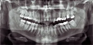
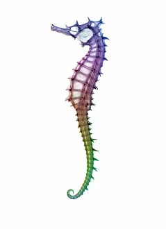
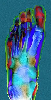
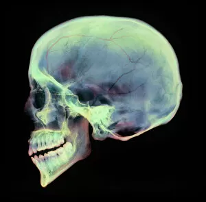
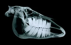
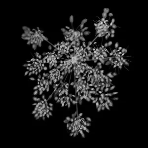
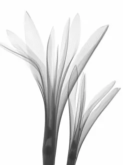
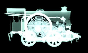
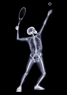
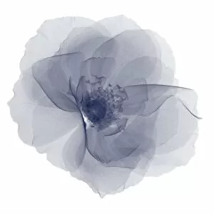
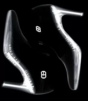
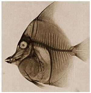
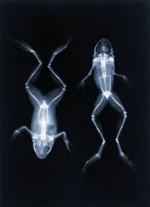
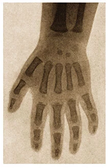
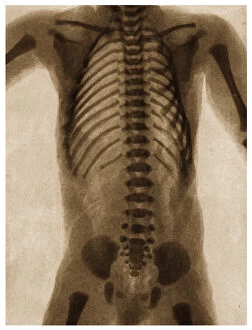
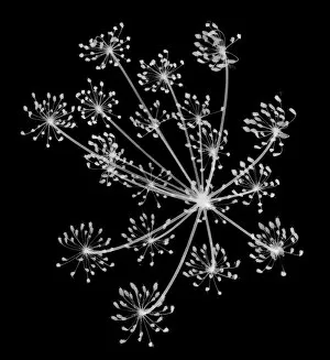
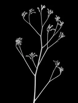
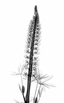
"Unveiling the Hidden World: Exploring Radiographs Beyond the Ordinary" Step into a captivating realm where radiographs reveal secrets hidden beneath our very eyes. From panoramic dental X-rays capturing intricate details of teeth to seahorse skeletons showcasing nature's delicate beauty, these images offer a glimpse into the mesmerizing world of radiography. Delving deeper, we encounter an ordinary foot transformed by an extraordinary X-ray, exposing its inner structure with remarkable precision. A human skull emerges from darkness, unveiling its intricate contours and reminding us of our mortality. Meanwhile, a horse skull stands as a testament to strength and grace in both life and death. But it doesn't stop there - bunions caught on an X-ray remind us that even seemingly small imperfections can leave their mark. In another twist, irregular heartbeats are captured through this powerful imaging technique, shedding light on conditions that affect millions worldwide. Shifting gears towards artistry within science, we witness an MRI-style X-ray revealing a leg adorned in stiletto shoes – merging fashion with medical technology in stunning detail. Parsley springs to life under the gaze of radiographic vision while tulips and gladiolus showcase their vibrant petals through translucent layers. And finally, behold the Oriental stargazer lily – its ethereal beauty enhanced by colored X-rays that transform it into a work of art beyond imagination. Radiographs have transcended mere diagnostic tools; they have become windows into worlds unseen. As we explore these captivating images together, let us marvel at the wonders revealed by this incredible fusion of science and artistry.