
Medulla Collection
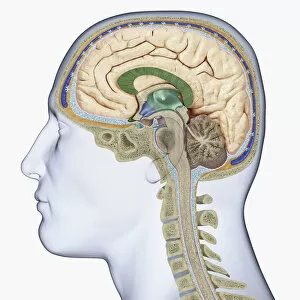
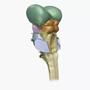
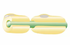
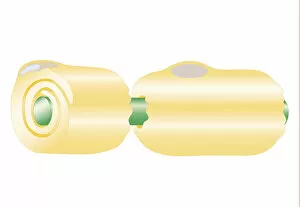
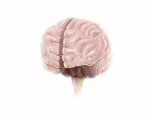
The medulla, an essential part of the human brain, can be observed from various angles and perspectives


All Professionally Made to Order for Quick Shipping
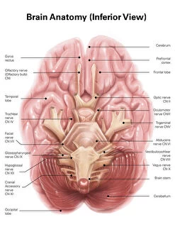
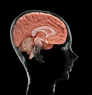
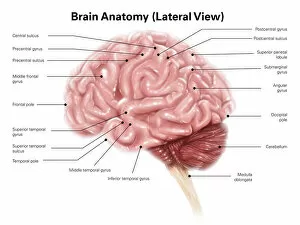
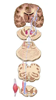
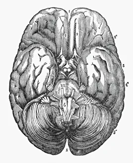
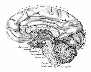
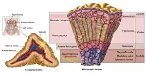
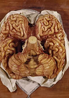
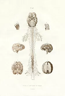
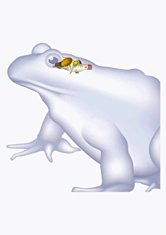
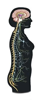
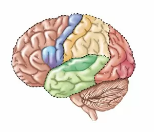
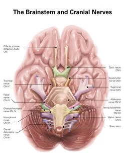
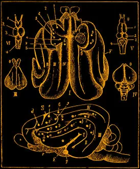
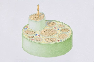
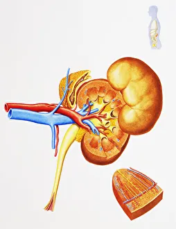
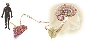
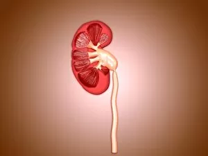
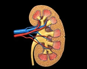
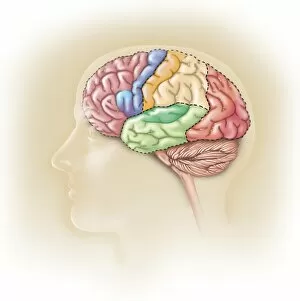
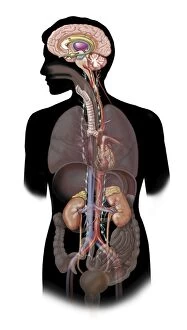
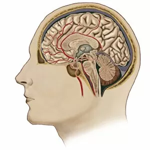
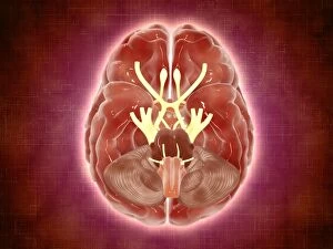
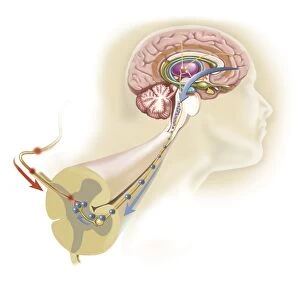
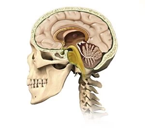
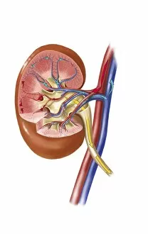
The medulla, an essential part of the human brain, can be observed from various angles and perspectives. From an inferior view, one can witness its intricate placement within the anatomy of the brain. A model of the human brain showcases this vital structure alongside other components like the cerebrum and cerebellum. When viewed laterally, the medulla's position becomes clearer in relation to other regions such as the motor cortex pathways. An engraved illustration from 1880 provides a glimpse into the under surface of the human brain, where one can appreciate both its complexity and beauty. Comparatively, even bird brains possess a medulla among their cerebral structures as depicted in another illustration. Artwork dedicated solely to exploring brain anatomy further highlights this crucial region's significance. In contrast, artwork showcasing kidneys reminds us that while different organs serve distinct purposes within our bodies, they all rely on proper communication with the nervous system facilitated by structures like the medulla. Scientific illustrations depicting side views offer valuable insights into how our brains are organized and interconnected. A wax model further aids in visualizing this complex organ's composition and function. Dating back to 1833-39, a colored engraving illustrates not only aspects of our nervous system but also emphasizes key elements such as pons and pineal gland alongside none other than our trusty medulla oblongata. Whether through anatomical models or artistic representations spanning centuries, exploring various depictions of "medulla" sheds light on its role within our intricate neural networks – reminding us that understanding these inner workings is fundamental for comprehending ourselves better.