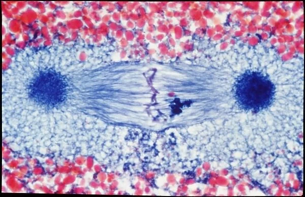
LM of metaphase mitosis in a triton (sea slug) egg
![]()

Wall Art and Photo Gifts from Science Photo Library
LM of metaphase mitosis in a triton (sea slug) egg
Metaphase cell division. Light micrograph of cell division (mitosis) in a triton (naked sea slug/ nudibranch) egg. This is the metaphase stage in mitosis. The spindles (blue horizontal threads) connecting the centrioles (dark blue ovals at centre left and right) are fully formed. The chromosomes (dark blue threads of DNA) have lined up on the spindles (centre) prior to division between the two potential daughter cells. Mitosis is a 5 stage process of cell division; it has two functions: replicated genetic material is distributed between the two potential cells, and the cell is cleaved in two. Magnification: x200 at 5x7cm size. x100 at 35mm
Science Photo Library features Science and Medical images including photos and illustrations
Media ID 6454133
© CNRI/SCIENCE PHOTO LIBRARY
Cell Division Histology Metaphase Mitosis Sea Slug Triton Light Micrograph
EDITORS COMMENTS
This print showcases the intricate process of metaphase cell division in a triton (sea slug) egg. The image, captured using a light microscope, provides a mesmerizing view into the world of mitosis. At this specific stage, known as metaphase, the spindles can be seen connecting the centrioles located at the center left and right of the frame. These fully formed spindles serve as crucial structures for dividing genetic material. The chromosomes, depicted as dark blue threads of DNA, have aligned themselves along these spindles in preparation for their eventual separation between two potential daughter cells. Mitosis itself is an elaborate five-stage process that not only ensures equal distribution of replicated genetic material but also results in cleavage of the cell into two distinct entities. With its magnification set at x200 and printed on a 5x7cm surface or x100 on 35mm film, this stunning photograph offers viewers an up-close look at animal cell division within a triton's egg. Its scientific significance lies in shedding light on fundamental biological processes occurring within organisms like sea slugs while simultaneously showcasing the beauty inherent in microscopic worlds.
MADE IN THE UK
Safe Shipping with 30 Day Money Back Guarantee
FREE PERSONALISATION*
We are proud to offer a range of customisation features including Personalised Captions, Color Filters and Picture Zoom Tools
SECURE PAYMENTS
We happily accept a wide range of payment options so you can pay for the things you need in the way that is most convenient for you
* Options may vary by product and licensing agreement. Zoomed Pictures can be adjusted in the Basket.

