Fraud Blocker Skip to main content

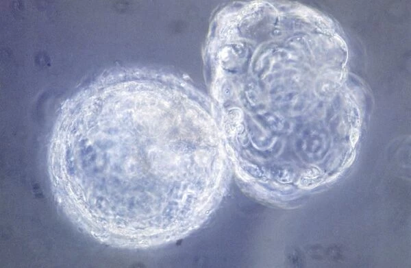
LM of hatching blastocyst in IVF

LM of hatching blastocyst in IVF
In Stock
We accept all major payment options so you can pay for the things you need in the way that is most convenient for you
Framed Photos
Buy from £59.99
Photo Prints
Buy from £6.99
Jigsaw Puzzles
Buy from £32.99
Poster Prints
Buy from £16.99
Canvas Prints
Buy from £34.99
Fine Art Prints
Buy from £49.99
Mounted Prints
Buy from £17.99
Metal Prints
Buy from £79.99
Cards
Buy from £8.97
Home Decor
Buy from £11.99

Science Photo Library Photo Prints and Wall Art

LM of hatching blastocyst in IVF

Light micrograph showing a hatching blastocyst, a six days-old human embryo. The blastocyst (left) at six days post-fertilisation is a hollow ball of cells with a fluid centre. Most of these cells will form the placenta & membranes, & only a small group (the inner cell mass) form the embryo proper. Here, the blastocyst is seen emerging from a hole in the zona pellucida (a protein " shell" around the oocyte). At this stage in nature, the blastocyst would be in the uterus & ready to begin implanting into the endometrium (the uterine lining). This image was produced in the course of In Vitro fertilisation research. Science Photo Library features Science and Medical images including photos and illustrations. © ANDY WALKER, MIDLAND FERTILITY SERVICES/ SCIENCE PHOTO LIBRARY

Media ID 6455615

Blastocyst Embryo Hatching In Vitro Fertilisation Light Micrograph

SECURE PAYMENTS
We accept all major payment options so you can pay for the things you need in the way that is most convenient for you

Made in UK
Safe Shipping, Easy Returns

FREE PERSONALISATION
We are proud to offer a range of customisation including Cropping, Captions, and Filters. Zoom Cropping can be adjusted in the basket


EDITORS COMMENTS

This print captures the intricate beauty of a hatching blastocyst, providing a glimpse into the early stages of human life. Taken using a light micrograph, this image showcases a six-day-old embryo formed through in vitro fertilization (IVF). The blastocyst, depicted on the left side of the frame, appears as a delicate hollow ball composed of cells surrounding a fluid-filled center. Within this remarkable structure, most cells will go on to develop into crucial components such as the placenta and membranes. Only a small group known as the inner cell mass holds the potential to form the actual embryo itself. Here, we witness an extraordinary moment as the blastocyst emerges from an opening in its protective protein shell called zona pellucida. In nature's course, at this stage, it would be within the uterus and prepared for implantation into the endometrium –the lining that nurtures pregnancy. This particular image was captured during research conducted in IVF laboratories where scientists strive to understand and enhance fertility treatments. The Science Photo Library presents this awe-inspiring photograph not for commercial purposes but rather to showcase scientific advancements and provide insight into our understanding of embryonic development. It serves as both an educational tool and testament to humanity's ongoing quest for knowledge about reproduction and early human life.


Framed Prints of LM of hatching blastocyst in IVF

Discover the wonders of life's earliest stages with Media Storehouse's Framed Prints featuring this captivating image from Science Photo Library. Witness the awe-inspiring moment of a hatching blastocyst, a six-day-old human embryo, as it transforms from a hollow ball of cells into a developing organism. This light micrograph showcases the intricate details of this vital stage in the in-vitro fertilization process, making it an inspiring addition to any office or home décor. Experience the beauty and complexity of life through the lens of science with Media Storehouse's Framed Prints.


Photo Prints of LM of hatching blastocyst in IVF

Discover the marvels of human development with Media Storehouse's Photographic Prints featuring this captivating image from Science Photo Library. Witness the miracle of life as a hatching blastocyst, a six-day-old human embryo, emerges from its protective shell in this stunning light micrograph. Each print is meticulously crafted to bring the intricacies of science to your home or office, igniting curiosity and inspiring wonder. Order yours today and bring the beauty of science into your space.


Poster Prints of LM of hatching blastocyst in IVF

Discover the wonders of human development with our Media Storehouse Poster Prints featuring the captivating image of a hatching blastocyst. This stunning light micrograph from Science Photo Library showcases a six-day-old human embryo, where the blastocyst transforms from a compact ball of cells into an expanding structure. Bring this remarkable moment in IVF science into your home or office with our high-quality, vibrant poster prints, perfect for inspiring curiosity and igniting conversations.


Jigsaw Puzzles of LM of hatching blastocyst in IVF

Discover the wonders of life's earliest stages with Media Storehouse's Jigsaw Puzzles. This intriguing puzzle, featuring a captivating image from Science Photo Library, showcases a hatching blastocyst in the process of in-vitro fertilization (IVF). Delve into the microscopic world and piece together this six-day-old human embryo, a hollow ball of cells with a fluid centre, to gain a deeper understanding of the marvels of life's beginnings. Engage your mind and bring this scientific marvel to life with our high-quality, challenging puzzles.

diamond
Made With High Grade Materials
thumb_up
Professional Quality Finish
package_2
Carefully Packed To Aid Safe Arrival

You May Also Like


©EyeUbiquitous_20008327©EyeUbiquitous_20008327
Add to Basket
Manuscript of Kenilworth, first published in 1821. Artist: Sir Walter ScottManuscript of Kenilworth, first published in 1821. Artist: Sir Walter Scott
Add to Basket
Standard of the Life-Guards Horse Regiment, 1832. Artist: Flags, Banners and StandardsStandard of the Life-Guards Horse Regiment, 1832. Artist: Flags, Banners and Standards
Add to Basket
The medicine, c1905The medicine, c1905
Add to Basket
A street in Flushing, Cornwall. Early 1900sA street in Flushing, Cornwall. Early 1900s
Add to Basket
Hungary, Budapest, Parliament and Danube RiverHungary, Budapest, Parliament and Danube River
Add to Basket
Spain, Canary Islands, Tenerife, Puerto de la Cruz, dawn view of Pico del TeideSpain, Canary Islands, Tenerife, Puerto de la Cruz, dawn view of Pico del Teide
Add to Basket
Caltagirone, Sicily, Italy, Mediterranean, EuropeCaltagirone, Sicily, Italy, Mediterranean, Europe
Add to Basket
INDIA Rajasthan Pushkar Cattle and camels brought to water in evening during fairINDIA Rajasthan Pushkar Cattle and camels brought to water in evening during fair
Add to Basket
WEST INDIES Barbados St James Angry Annies brightly decorated traditional Barbadian food restaurant and bar in HoletownWEST INDIES Barbados St James Angry Annies brightly decorated traditional Barbadian food restaurant and bar in Holetown
Add to Basket
Constantine the Great, Roman EmperorConstantine the Great, Roman Emperor
Add to Basket
Cityscape at sunset, Staithes, Yorkshire, EnglandCityscape at sunset, Staithes, Yorkshire, England
Add to Basket

Gallery Wall Inspiration

Sensory homunculus
Plague doctor, 17th century artwork
Brain pathways
Brain blood vessels, 3D angiogram C007 / 1981
Sensory homunculus
Cerebellum tissue, light micrograph
Purkinje nerve cells in the cerebellum
Brain fibres, DTI MRI scan C017 / 7035
Medulla oblongata in the brain, artwork
Nerve and glial cells, light micrograph
Motor and sensory homunculi
Nerve cell, SEM
Phrenological model of personality traits
Diagram of the human brain and spinal column
Phrenological Head / Scrap
Nerve cell
Moth antenna, SEM
Touch receptors
Anatomy of the human bloodstream, lithograph, published in 1874
"Manos de Artista". The hands of a young man illuminated by the bright light
Face of a young woman from Taormina
Statue of Antinous-Osiris or Egyptian Apollo, Gregorian Egyptian Museum, Vatican Museums, Vatican City
Motor homunculus model
Motor homunculus
Brain fibres, DTI MRI scan C017 / 7099
Normal knees, X-ray
The Return of the Prodigal Son, c1668. Artist: Rembrandt Harmensz van Rijn


© Copyright Media Storehouse, All Rights Reserved 2003 - 2024
Facebook     Pinterest     100% Payment Secure     Reviews IO