Home > Science > SEM
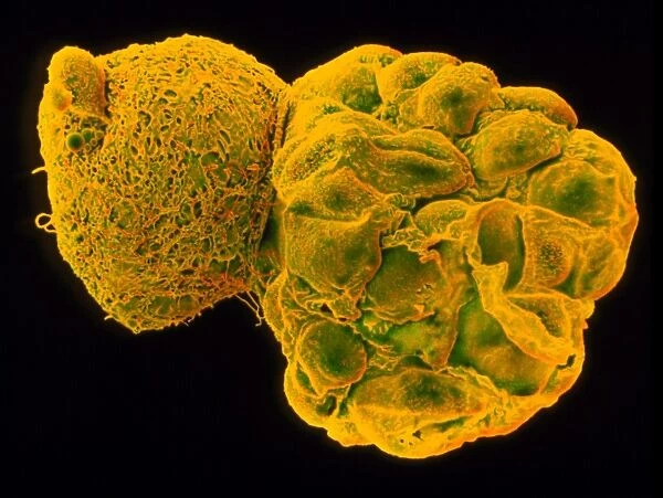
Coloured SEM of a hatching blastocyst 5 days old
![]()

Wall Art and Photo Gifts from Science Photo Library
Coloured SEM of a hatching blastocyst 5 days old
Hatching blastocyst embryo. Coloured scanning electron micrograph (SEM) of a human embryo at the blastocyst stage, five days after fertilisation. It is seen at right hatching from a hole in the zona pellucida (at left), a protein shell that originally surrounded the unfertilised egg. The blastocyst is a hollow ball of cells with a fluid centre. Each cell is called a blastomere. Most of these embryo cells will form the placenta and membranes around the embryo, and only a small group (the inner mass) form the embryo proper. At this stage the blastocyst has moved into the uterus (womb) and is preparing to implant on the womb wall. Magnification: x360 at 6x7cm size. Magnification; x1, 200 at 8x10 inch size
Science Photo Library features Science and Medical images including photos and illustrations
Media ID 6453841
© DR YORGOS NIKAS/SCIENCE PHOTO LIBRARY
FEATURES IN THESE COLLECTIONS
EDITORS COMMENTS
This print showcases the intricate beauty of life at its earliest stages. Captured through a scanning electron microscope, it features a coloured SEM image of a hatching blastocyst embryo that is five days old. The blastocyst, seen on the right side of the image, is emerging from a small opening in the zona pellucida—a protective protein shell that once enveloped the unfertilised egg. The blastocyst itself resembles a hollow ball composed of numerous cells known as blastomeres. Remarkably, most of these cells will go on to form vital components such as the placenta and membranes surrounding the developing embryo. Only a select group called the inner mass will eventually give rise to what we recognize as an embryo. At this critical stage, approximately five days after fertilisation has taken place, the blastocyst has migrated into the uterus and is preparing for implantation onto its wall—an essential step in pregnancy initiation. With magnification levels reaching x360 at 6x7cm size or x1,200 at 8x10 inch size, this visually stunning photograph offers us an extraordinary glimpse into our own beginnings—the miraculous journey from conception to new life.
MADE IN THE UK
Safe Shipping with 30 Day Money Back Guarantee
FREE PERSONALISATION*
We are proud to offer a range of customisation features including Personalised Captions, Color Filters and Picture Zoom Tools
SECURE PAYMENTS
We happily accept a wide range of payment options so you can pay for the things you need in the way that is most convenient for you
* Options may vary by product and licensing agreement. Zoomed Pictures can be adjusted in the Basket.

