Photographic Print : Colour SEM of seminiferous tubule of the testis

![]()
Photo Prints from Science Photo Library
Colour SEM of seminiferous tubule of the testis
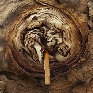
Seminiferous tubule. Coloured Scanning Electron Micrograph (SEM) of a sectioned seminiferous tubule, the site of sperm production in the human testis. The tubule contains a swirl of forming sperm cells (blue). Each testis is packed with seminiferous tubules. The tubules are lined by a stratified epithelium containing two types of cell: Sertoli cells, which nourish developing sperm, and spermatogenic cells, which produce the sperm. The sperm are released into the cavity of the tubule to migrate to the epididymis, where they mature. Around the seminiferous tubule are Leydig cells (orange) which produce testosterone. Magnification: x200 at 5x7cm size
Science Photo Library features Science and Medical images including photos and illustrations
Media ID 6451259
© CNRI/SCIENCE PHOTO LIBRARY
Re Production Reproductive System Seminiferous Tubule Site Sperm Spermatogenesis Spermiogenesis Testis
10"x8" (25x20cm) Photo Print
Discover the intricacies of the human body with our Media Storehouse range of Photographic Prints. This captivating image showcases a Coloured Scanning Electron Micrograph (SEM) of a sectioned seminiferous tubule, revealing the complex structure of the site of sperm production in the testis. Bring the wonders of science into your home or office with our high-quality, vibrant prints. Each print is meticulously produced to preserve the stunning detail and clarity of the original image, making it a must-have for any science enthusiast or educational setting.
Printed on archival quality paper for unrivalled stable artwork permanence and brilliant colour reproduction with accurate colour rendition and smooth tones. Printed on professional 234gsm Fujifilm Crystal Archive DP II paper. 10x8 for landscape images, 8x10 for portrait images.
Our Photo Prints are in a large range of sizes and are printed on Archival Quality Paper for excellent colour reproduction and longevity. They are ideal for framing (our Framed Prints use these) at a reasonable cost. Alternatives include cheaper Poster Prints and higher quality Fine Art Paper, the choice of which is largely dependant on your budget.
Estimated Image Size (if not cropped) is 18.2cm x 25.4cm (7.2" x 10")
Estimated Product Size is 20.3cm x 25.4cm (8" x 10")
These are individually made so all sizes are approximate
Artwork printed orientated as per the preview above, with portrait (vertical) orientation to match the source image.
EDITORS COMMENTS
This print showcases a mesmerizing view of the seminiferous tubule within the testis. In this coloured Scanning Electron Micrograph (SEM), we are granted a glimpse into the intricate process of sperm production in the human body. The seminiferous tubule, depicted here, is teeming with activity as forming sperm cells take shape and develop. The testis, an organ vital to reproduction, houses numerous seminiferous tubules like these. These tubules are lined by a stratified epithelium consisting of two types of cells: Sertoli cells and spermatogenic cells. While Sertoli cells provide nourishment for developing sperm, spermatogenic cells play a crucial role in producing them. As we observe closely, we can witness a swirling mass of blue-coloured spermatozoa within the tubule's cavity. Once matured, these remarkable sperm will be released into the epididymis to continue their journey towards fertilization. Surrounding the seminiferous tubule are Leydig cells portrayed in vibrant orange hues. These specialized cells hold another important responsibility – they produce testosterone which plays a pivotal role in male reproductive functions. With magnification at x200 and presented on a 5x7cm size print, this image from Science Photo Library offers us an awe-inspiring insight into one of nature's most extraordinary processes - spermiogenesis. It serves as both an educational tool and an artistic representation that celebrates the wonders hidden
MADE IN THE UK
Safe Shipping with 30 Day Money Back Guarantee
FREE PERSONALISATION*
We are proud to offer a range of customisation features including Personalised Captions, Color Filters and Picture Zoom Tools
SECURE PAYMENTS
We happily accept a wide range of payment options so you can pay for the things you need in the way that is most convenient for you
* Options may vary by product and licensing agreement. Zoomed Pictures can be adjusted in the Basket.