Mounted Print : Scanning electron micrograph of cell division
![]()

Mounted Prints from Science Photo Library
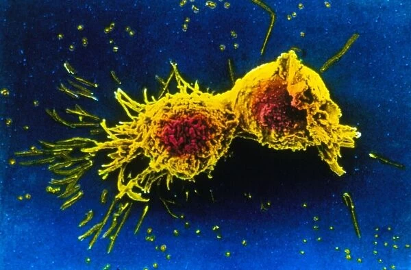
Scanning electron micrograph of cell division
Cell division. Coloured scanning electron micrograph of two human cells pulling apart in the final stage or telophase of cell division. Cell division (cytokinesis) follows mitosis, where the nucleus separates into two, each containing an identical set of chromosomes. Magnification x2000 at 7x5cm size. Magnification x1000 at 35mm size
Science Photo Library features Science and Medical images including photos and illustrations
Media ID 6455227
© CNRI/SCIENCE PHOTO LIBRARY
Cell Division Cytokinesis Mitosis Telophase
10"x8" Mount with 8"x6" Print
Discover the intricacy of life with our Media Storehouse Mounted Photos. This captivating image showcases the final stage of cell division, known as telophase. Witness the breathtaking detail of two human cells pulling apart, as revealed in this coloured scanning electron micrograph from Science Photo Library. Each mounted photo is meticulously printed on high-quality photographic paper and mounted on a sturdy backing, ensuring a lasting impression. Bring the wonders of science into your home or office with our exquisite range of mounted photos.
Printed on 8"x6" paper and suitable for use in a 10"x8" frame (frame not included). Prints are mounted with card both front and back. Featuring a custom cut aperture to match chosen image. Professional 234gsm Fujifilm Crystal Archive DP II paper.
Photo prints supplied in custom cut card mount ready for framing
Estimated Image Size (if not cropped) is 20.3cm x 13.3cm (8" x 5.2")
Estimated Product Size is 25.4cm x 20.3cm (10" x 8")
These are individually made so all sizes are approximate
Artwork printed orientated as per the preview above, with landscape (horizontal) orientation to match the source image.
EDITORS COMMENTS
This print showcases the intricate process of cell division in stunning detail. In this scanning electron micrograph, we witness two human cells reaching the final stage of telophase, where they begin to separate from each other. The vibrant colors used to enhance the image highlight the significance and complexity of cytokinesis, which follows mitosis. During mitosis, the nucleus splits into two identical sets of chromosomes within each cell. As depicted in this photograph, these dividing cells are pulling apart as part of their natural progression towards creating new life. With a magnification level of x2000 at a 7x5cm size or x1000 at 35mm size, every tiny detail is brought to life with astonishing clarity. The sheer beauty and intricacy captured by Science Photo Library's talented photographers remind us of the wonders that occur within our own bodies on a microscopic level. This image serves as a reminder that even seemingly small processes like cell division play an essential role in maintaining our overall health and well-being.
MADE IN THE UK
Safe Shipping with 30 Day Money Back Guarantee
FREE PERSONALISATION*
We are proud to offer a range of customisation features including Personalised Captions, Color Filters and Picture Zoom Tools
SECURE PAYMENTS
We happily accept a wide range of payment options so you can pay for the things you need in the way that is most convenient for you
* Options may vary by product and licensing agreement. Zoomed Pictures can be adjusted in the Basket.


