Jigsaw Puzzle : Flower reproductive parts, SEM


![]()
Jigsaw Puzzles from Science Photo Library
Flower reproductive parts, SEM
Flower reproductive parts. Coloured SEM (scanning electron micrograph) of the reproductive parts of an unidentified flower. The smooth structure that runs diagonally from lower right to upper left is the upper part of the pistil (female reproductive structure). Seen here is the style (stalk-like) that supports the stigma at its tip. The ovary is not seen as it lies at the base of the pistil. The style is surrounded by the pollen sacs (anthers, male reproductive structures). The protective case that enclosed these structures before the flower opened is at upper left. Magnification unknown
Science Photo Library features Science and Medical images including photos and illustrations
Media ID 6287551
© SUSUMU NISHINAGA/SCIENCE PHOTO LIBRARY
Anther Anthers Carpel Carpels Cluster Filament Filaments Flowering Part Group Parts Pistil Pollen Re Production Reproductive Structures
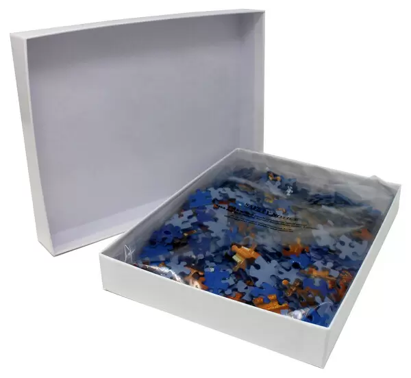
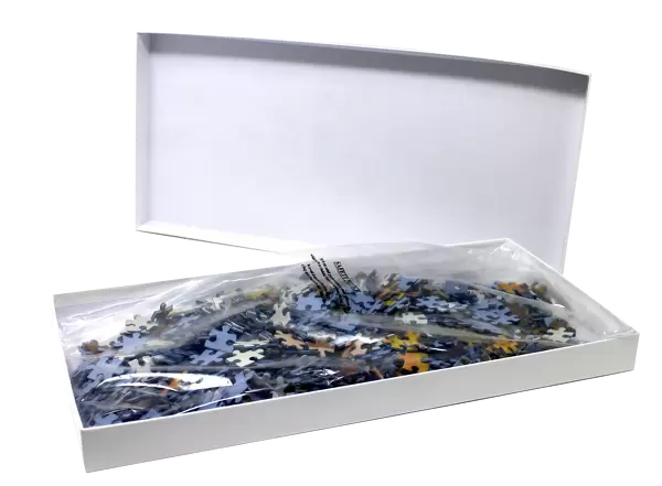
Jigsaw Puzzle (500 Pieces)
Explore the intricacies of nature with our Media Storehouse Jigsaw Puzzles, featuring the captivating image "Flower reproductive parts, SEM" from Science Photo Library. Delve into the world of science and botany as you piece together this intricately detailed, coloured scanning electron micrograph (SEM) of unidentified flower reproductive parts. This puzzle is not only a fun and engaging activity but also an educational experience, perfect for those with a passion for science, nature, or just a love for puzzles. Engage your mind and challenge yourself to uncover the hidden beauty within this complex floral arrangement.
500 piece puzzles are custom made in the UK and hand-finished on 100% recycled 1.5 mm millboard. There is a level of repetition in jigsaw shapes with each matching piece away from its pair. The completed puzzle measures 38x50cm and is delivered packaged in an attractive presentation box specially designed to fit most letter box slots
Jigsaw Puzzles are an ideal gift for any occasion
Estimated Product Size is 38cm x 50.2cm (15" x 19.8")
These are individually made so all sizes are approximate
Artwork printed orientated as per the preview above, with landscape (horizontal) or portrait (vertical) orientation to match the source image.
EDITORS COMMENTS
This print showcases the intricate beauty of flower reproductive parts. Taken using a scanning electron microscope (SEM), this coloured SEM image reveals the fascinating details of an unidentified flower's reproductive structures. The smooth structure running diagonally from lower right to upper left is the upper part of the pistil, which serves as the female reproductive organ. Specifically, we can observe the style, a stalk-like structure that supports the stigma at its tip. Although not visible in this image, the ovary lies at the base of the pistil. Surrounding and contrasting with the pistil are multiple pollen sacs known as anthers, representing male reproductive structures. These anthers enclose precious pollen grains responsible for fertilization. At upper left, we can also see remnants of a protective case that once enclosed these delicate structures before they bloomed into full glory. The magnification level used to capture this mesmerizing view remains unknown but undoubtedly highlights nature's incredible attention to detail. This photograph offers botany enthusiasts and curious minds alike a glimpse into plant anatomy and reproduction processes while showcasing both male and female components harmoniously existing within one flower.
MADE IN THE UK
Safe Shipping with 30 Day Money Back Guarantee
FREE PERSONALISATION*
We are proud to offer a range of customisation features including Personalised Captions, Color Filters and Picture Zoom Tools
SECURE PAYMENTS
We happily accept a wide range of payment options so you can pay for the things you need in the way that is most convenient for you
* Options may vary by product and licensing agreement. Zoomed Pictures can be adjusted in the Basket.