Jigsaw Puzzle > Popular Themes > Human Body
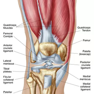
Jigsaw Puzzle : Coloured X-ray of a human knee joint


![]()
Jigsaw Puzzles from Science Photo Library
Coloured X-ray of a human knee joint
Knee joint. Coloured X-ray of a healthy human knee joint. Two bones meet at the knee forming a joint that works like a hinge. At top is the large femur (thigh-bone), which articulates with the tibia (shin-bone) at bottom. Next to the tibia (at lower right) is the smaller fibula bone. The patella or kneecap (above centre, faint blue oval) is a protective bone at the front of the knee held in position by muscles and tendons. Two discs of protective cartilage cover the surfaces of the femur and tibia to reduce friction between these bones. This joint, the largest in the body, allows a backward-forward hinge movement with slight rotation
Science Photo Library features Science and Medical images including photos and illustrations
Media ID 6419970
© MEHAU KULYK/SCIENCE PHOTO LIBRARY
Bones Femur Joint Knee Knee Cap Knee Joint Patella Tibia Hinge Joint
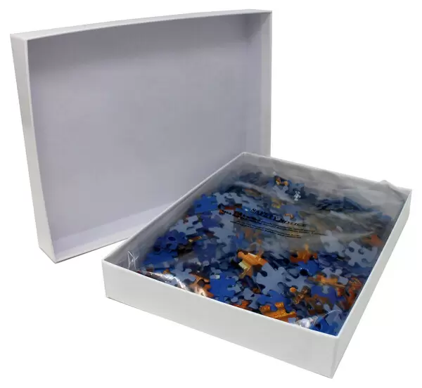
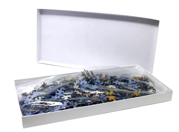
Jigsaw Puzzle (400 Pieces)
Discover the intricacies of the human body with our captivating Coloured X-ray Jigsaw Puzzle from Media Storehouse. Featuring an image of a healthy human knee joint by Science Photo Library, this puzzle presents a unique and educational challenge. Piece together the intricate details of the knee's bones and their alignment, revealing the complex mechanism that allows us to bend and straighten our legs. A must-have for puzzle enthusiasts, science lovers, and anyone seeking a brain-teasing activity. Engage your mind and unlock the mysteries of the body, one puzzle piece at a time.
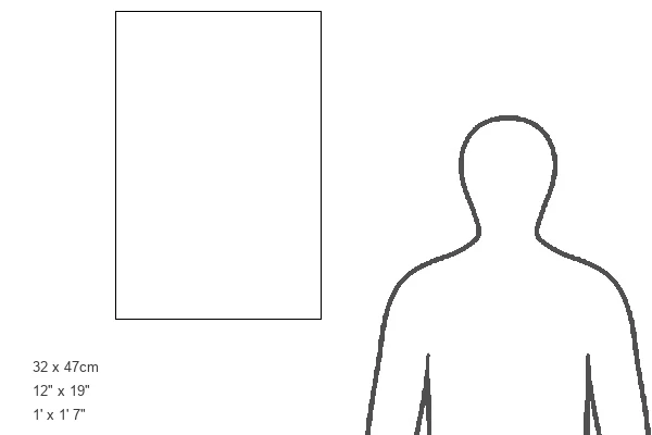
400 piece puzzles are custom made in the UK and hand-finished on 100% recycled 1.5 mm millboard. There is a level of repetition in jigsaw shapes with each matching piece away from its pair. The completed puzzle measures 31x47cm and is delivered packaged in an attractive presentation box specially designed to fit most letter box slots
Jigsaw Puzzles are an ideal gift for any occasion
Estimated Product Size is 31.5cm x 47.2cm (12.4" x 18.6")
These are individually made so all sizes are approximate
Artwork printed orientated as per the preview above, with portrait (vertical) orientation to match the source image.
EDITORS COMMENTS
This print showcases a coloured X-ray of a healthy human knee joint, revealing the intricate anatomy and functionality of this crucial body part. At first glance, it becomes evident that two bones converge at the knee to form a remarkable hinge-like joint. The larger femur, also known as the thigh-bone, sits atop and artfully connects with the tibia or shin-bone at the bottom. Adjacent to the tibia lies its smaller companion, the fibula bone. A faint blue oval shape positioned above center steals our attention - this is none other than the patella or kneecap. Serving as a protective shield for the front of our knee, it remains securely in place thanks to surrounding muscles and tendons. To ensure smooth movement between these bones, two discs composed of protective cartilage cover their surfaces. This ingenious design reduces friction and enables seamless motion within this largest joint in our entire body. The knee joint's unique construction allows for an impressive backward-forward hinge movement with slight rotation capabilities. Its significance cannot be overstated; every step we take relies on its strength and flexibility. Through this mesmerizing image captured by Science Photo Library, we gain profound insight into our own skeletal structure while appreciating both its complexity and elegance simultaneously.
MADE IN THE UK
Safe Shipping with 30 Day Money Back Guarantee
FREE PERSONALISATION*
We are proud to offer a range of customisation features including Personalised Captions, Color Filters and Picture Zoom Tools
SECURE PAYMENTS
We happily accept a wide range of payment options so you can pay for the things you need in the way that is most convenient for you
* Options may vary by product and licensing agreement. Zoomed Pictures can be adjusted in the Basket.